Claude Franceschi
AbstractA clear understanding of the physical concepts of the venous hemodynamics improves the knowledge, diagnosis and treatment of the venous insufficiency. For this reason they will be presented, defined and weighted according to their physiologic and pathologic incidence.
|
Represented by a vector, the Force is the magnitude, direction and acceleration of the interaction between two physical bodies. This interaction can be attractive or repulsive. It increases with the rate of change of velocity (v) called acceleration (a), and with the quantity of inertia of the object called mass (m).
F=ma
Acceleration unit (a) is m/s2 (meters per second squared).
Force unit is the Newton (N), i.e. the force that applied to a mass of 1 kg produces an acceleration of 1 m/s2.
In addition and in an operative manner, Force can be also defined as an entity, which can be measured by a dynamometer.
The two definitions are equivalent.
Mechanical Energy is the capacity of a physical system to perform work, i.e. the integral of the force over a distance of displacement (Newton × meter or Joule). It can be expressed in several forms such as heat, electricity, light, mechanical energy, potential energy, kinetic energy, etc.
The Principle of Conservation of Energy states that energy is neither created nor destroyed but can only be transformed from one form to another in an isolated system.
In venous hemodynamics, the energy is mainly expressed or converted into the forms of potential and kinetic energy.
It is called Potential Energy (P.E.) or stored energy when it refers to the ability of a system to do work due to its position such as Gravitational Potential Energy (G.P.E) stored along the vertical vector of the gravitational acceleration (height) or in internal structure such as Elastic Potential Energy is energy stored in a spring.
It is called Kinetic Energy (K.E.) when it represents the energy of a mass (m) in motion velocity (v). It is a scalar quantity therefore always positive with the same work units such as Joules (J) Newton × m.
K.E.=(1/2) mv2
According to the Principle of Conservation of Energy, P.E. and K.E. being two expressions of the same energy they can be converted between each other while the value of the total energy (T.E.) remains constant.
TE=P.E+K.E=Konst
The venous pressure of the lower limbs is made of three main components that vary independently: the Gravitational Hydrostatic Pressure, the Residual Pressure and the Muscular and Respiratory Pressure, which result from the energy of Gravity and pumps (Cardiac, Respiratory and Muscular).
Pressure (P) is Force (F) exerted per unit area (S): P=F/S. The international unit is the Pascal (Pa), which is 1 N/m2
1 Torr=133.3224 Pa=1 mmHg=1.35 cm H2O.
In fluid mechanics, Pressure is expressed according to the form of energy, the direction and rate of force, the physical features of the fluid and the solid structures that interact. Due to the complexity of the fluid mechanics many equations and laws are described. The main ones are Newton, Pascal, Bernouilli, Poiseuille, Reynolds, Navier-Stokes equations. Each one relates to a particular aspect of the issue and limited to few and strict initial conditions therefore none of them can measure very accurately the pressure and its various expressions. In practice their approximate values are most of the time sufficient for the understanding and the assessment of hemodynamics, provided they are confirmed by experiment.
Initial conditions:
i) Points 1 (up-stream) and 2 (down-stream) lie on the stream line;
ii)
The fluid has constant density;
iii)
The flow is steady;
iv)
There is no friction.
Although these restrictions sound severe, the Bernoulli equation is very useful partly because of its simplicity to use and partly because it can give great insight into the balance between pressure, velocity and elevation (http://www.princeton.edu/~asmits/Bicycle_web/Bernoulli.html).
Total pressure=½+ρv2+ρgh+p=constant
V is the flow velocity, ρ is the fluid density (mass per unit of volume) and h is the elevation.
½ ρv2 is called the Dynamic Pressure (DP) because it arises from the motion of the fluid and relates to the Kinetic Energy. p is the Static Pressure (SP) because it increases with the slowing of the flow velocity and relates to part of the dynamic energy stored (converted) into Potential Energy according to the Law of Conservation of the Energy.
Notice that the energy DP+SP=Konst sometimes called stagnation pressure can be supplied by one or more sources (Gravity, the cardiac, respiratory and muscular pumps).
gh is the Hydrostatic Gravitational Pressure (HGP) that relates to potential energy of the fluid’s position, ρ density, h height is submitted to the gravitational acceleration g. Notice that HGP is not influenced by the flow velocity v. In the venous system, HGP is the most variable component of the venous pressure because it varies with the height of the blood column, particularly at the lower limbs extremities where the pressure changes at the ankle from negative when legs are elevated to 90 mmHg in a still upright position and around 20 mmHg when lying horizontally. This is the reason for the predominance of the venous insufficiency below the knee when this excessive HGP cannot be reduced in upright position.
Fortunately, the muscular activity (moving, walking, running, etc.) lessens it from 90 mmHg down to 30 mmHg thanks to the venous valves competence that fractionates the pressure column during the muscular contraction (systole) and relaxation (diastole) called Dynamic Fractioning of the HGP (DFHP). The DFHP impairment is the most frequent cause of the lower limbs venous insufficiency.
The pressure exerted by the fluid against the wall of the conduits is called Lateral Pressure (LP) or Parietal Pressure.
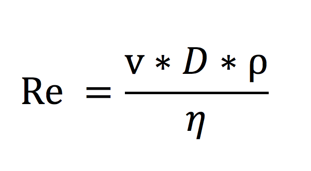
LP=SP+HGP when the flow is laminar, i.e. made of parallel layers without disruption. When a critical Reynolds number (Re) is exceeded the laminar flow changes into a turbulent one. When the flow is turbulent, layers are disrupted; the speed of the fluid at a given point is continuously undergoing changes in static pressure, magnitude and direction. Therefore turbulences are responsible for the pressure loss (driving pressure reduction) and for the corresponding converted energy into wall caloric/mechanical stress that could explain part of the wall changes (enlargement, thickening, varices, etc.) when veins are submitted to an excess of flow velocity.
Reynolds number:
v=velocity, D=vessel diameter, ρ=blood density, η=blood viscosity.
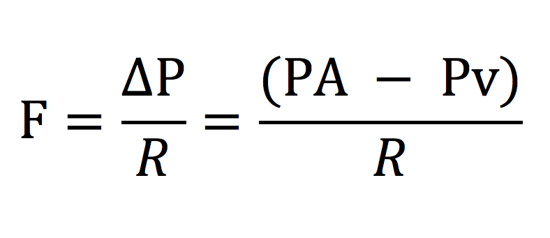
Regarding the hemodynamics, the Driving Pressure (DP) is generally the pressure difference (ΔP) between the arterial pressure (PA) and the venous pressure (Pv) referring to any point along the arterial flow and any other point along the venous flow. It can also be called Perfusion Pressure or Pressure Gradient. Flow (F) varies according to the Resistance (R) between the two chosen points.
The Resistance (R) in a conduit roughly obeys to the Poiseuille law where the radius (R) variation is greatly predominant to viscosity µ and length because of the fourth power r4.
R=µ*8L/(π*r4)
In the lower limbs, the main resistance to flow is due to the micro-circulation and varies with the microvascular caliber variations. At the venous end of the capillaries, the venous pressure supplied by the arterial pressure is called Residual Pressure (RP) while the gravitational hydrostatic pressure is due to the height of the venous blood column and varies independently.
ΔP=PA–Pv=F*R=F*µ*8L/(π*r4)
RP=Residual Pv=PA–F*R=PA–F*µ*8L/(π*r4)
Therefore, RP and Flow vary mainly with the modification of the microvascular vasomotion. RP is increased particularly by the heat (thermoregulation), inflammation and the muscular activity that dilate the microvascular vessels. A high diastolic flow due to low resistance is shown in daily practice by Doppler on the arterial side of the microcirculation. This phenomenon is maximal in arterio-venous fistulae (congenital, traumatic or therapeutic) where the RP may raise up to the arterial pressure and leads to signs of venous insufficiency.
RP increases also when a resistant obstacle blocks the venules and/or veins, because it slows the velocity of the microvessels where consequently the resistance decreases according to the Poiseuille law and leads to a reflex vasodilatation of the arterioles and micro-shunts opening. Whatever the mechanism, this increase of RP is attested by daily practice. This accounts for the venous tributary dilation in case of a venous blockage and is called Open Vicarious Shunts (OVS) because they are forced and overloaded by the consequent raise of RP, not only in the case of a deep venous obstacle but also when the superficial veins are occluded, e.g. after extensive flow ablation of superficial varicose veins. The venous claudication may occur while walking in the case of a venous obstacle because the excessive RP at rest is increased by the additional microvascular dilation and muscular pump pressure. The extreme effect of the downstream resistance excess can lead to an arterial flow stop responsible for ischemic phlegmasia cerulea.
The peripheral muscles act as an alternative pump (MP) such as the cardiac pump, i.e. contraction (systole) and relaxation (diastole) when the venous valves like the cardiac establish a unidirectional flow. Its action is limited to the action of walking, running, etc.). MP contributes to pumping the increased stress flow towards the heart and at the same time fractionates the GSP in order to reduce the venous pressure below the knee in an upright position. The energy supplied by the muscular activity is physiologically converted in a cardiopetal gradient pressure and flow. It can be more or less converted in cardiofugal gradient pressure and flow (reflux) whilst the DFHP is impaired according to the degree of venous valve incompetence. The flow of the superficial veins is not directly activated by the muscular pump (except for the Lejars foot sole pump) but may be aspirated through the perforators distally to the pump by the diastolic aspiration effect and proximally by Venturi’s effect during the systole.
What happens in the case of a superficial venous segment where all the valves are incompetent, from the deep-superficial junction (perforators, SFJ, SSPJ) proximal to the muscular pump down to a deep-superficial junction located distally to the muscular pump?
The valve incompetence makes the superficial reflux possible during the MP diastole that aspires back into the deep distal veins, part of the proximal deep venous flow through the proximal incompetent point (called escape point) then into the incompetent superficial segment and finally into the distal deep-superficial junction (called re-entry point whatever the competence of its valves). This phenomenon overloads its physiologic draining flow with deep flow that restitutes back part of the energy lifted up during the systole whilst the DFHP impairment does not permit a distal GHP fractioning. This closed circuit is called closed shunt (CS). Therefore the CSs are overloaded during the diastole of the MP.
In case of an obstacle, the OVS are overloaded by the systolic pressure of the MP.
Both systolic and diastolic effects of the MP on the magnitude and direction of the venous flow is easily assessed by US Duplex during rest and stress manoeuvers.
Squeezing/relaxing the calf is a usual method to test the venous competence but it is not properly physiologic such as the muscular pump activity in the manner that the cardiac massage reproduces the physiologic cardiac activity.
Some tests such as the Paranà manoeuver try to reproduce more closely the physiological body functioning.
During breathing, the diaphragm, thoracic, and abdominal muscles activates the variation of the venous volume of the inferior and superior cava vein and right atrium. The respiratory pump plays the role of an additional alternate pump, diastolic during the inspiration and systolic during the expiration. The cardiopetal venous gradient increases during the inspiration thanks to the thoracic volume increase while the abdominal cava vein is gently squeezed by the diaphragm descent.
In some extra respiratory circumstances like coughing, weight carrying, defecating, the conjoined contraction of the abdominal, thoracic and diaphragm muscles squeezes the intra-abdominal and intra-thoracic veins especially when the lung are full of air (i.e., at the end of a deep inspiration) and the expiration is blocked by glottis closure. This extreme blocked expiration increases the thoracoabdominal venous pressure leading to a reverse of the cardiopetal gradient of pressure into cardiofugal. The result is venous valve closure preventing the otherwise venous reflux. The test dedicated to detect the venous valve incompetence is called Valsalva manoeuver, especially from the proximal ones down to the popliteal vein. This manoeuver is mainly useful for the assessment of pelvis and SFJ escape points.
The cardiac pump supplies the RP during the left ventricle systole and the right ventricle increases the cardiopetal gradient of the venous pressure during the diastole. The increase of the venous pressure due to the right heart ventricle and valves impairment leading to distal edema is a regular diagnosis.
TMP is the key parameter of the venous hemodynamics in terms of tissue drainage and the volume and diameter of the venous compartment. It results from the hydrostatic difference between Intravascular Lateral pressure (IVLP) and the Extravascular pressure (EVP) which is the sum of the Atmospheric Pressure (Ath P) and the pressure of the surrounding tissues (Tis P).
Volume and pressure in a system change according to the system features. The venous bed, especially the large and Cava veins, shows a low variation of trans-mural pressure respect to the volume thanks a damn effect due to its capacitance (able to store 60-70% of the blood volume), its high passive compliance and the active tone control of the venous wall. This effect is sometimes called reservoir effect. The volume depends on the TMP, the active tone of the muscular media layer and the passive Compliance of the adventitial layer.
Notice that the HGP variations cannot be damped by the reservoir effect because HGP only depends on the height of the fluid and never on its volume (Pascal) in contrast with Static Pressure.
At the capillary level the drainage depends not only on TMP hydrostatic components (IVLP and EVP), but also on the osmotic gradient on either side of the capillary wall according to the Starling law. This is the reason why an excessive osmotic gradient can show the same signs (hampered drainage) as an excessive TMP when the protein rate is too low inside the plasma or excessive inside the interstitial fluids (macro-proteins trapped by lymphatic excessive pressure or lack of lymphatic vessels).
1. Klabunde RE. Cardiovascular physiology concepts. 2nd ed. Philadelphia, PA: Lippincott Williams & Wilkins; 2011.
2. Bar–Meir G. Basics of fluid mechanics (version 0.3.1.1). Available from: http://www.potto.org/ Accessed: December 21, 2011.
3. Tyberg JV. How changes in venous capacitance modulate cardiac output. Pflugers Arch 2002;445:10-7.[CrossRef][PubMed]
4. Franceschi C. Dynanamic fractionizing of hydrostatic pressure, closed and open shunts, vicarious varicose evolution: how these concepts made the treatment of varices evolve? Phlebologie 2003;56:61-6.
5. Franceschi C. La cure hemodynamique de l’insuffisance veineuse en ambulatoire. J Mal Vasc 1992;(17):291-300.[PubMed]
6. Franceschi C. Théorie et pratique de la cure conservatrice et hémodynamique de l’insuffisance veineuse en ambulatoire. Precy-sous-Thil: Editions de l’Armançon; 1988.
7. Franceschi C. Measures and interpretation of venous flow in stress tests. Manual compression and Parana manoeuver. Dynamic reflux index and Psatakis index. J Mal Vasc 1997;22:91–5.[PubMed]
[TOP]