|
|
|
Free Neuropathology 2:6 (2021) |
|
Review |
|
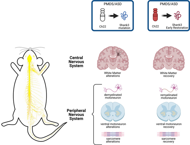
Neurodevelopmental disorders: 2021 update |
|
Alfonsa Zamora-Moratalla1, María Martínez de Lagrán1, Mara Dierssen1,2,3 |
|
1 Centre for Genomic Regulation (CRG), The Barcelona Institute of Science and Technology, Dr. Aiguader 88, Barcelona 08003, Spain |
|
Corresponding author: |
|
Submitted: 23 February 2021 Accepted: 12 March 2021 Copyedited by: Cathryn Cadwell Published: 24 March 2021 |
|
Keywords: Autism spectrum disorder, ASD, Neurodegenerative disorders, Next generation sequencing, Microbiome, Preterm birth |
|
Abstract One of the current challenges in the field of neurodevelopmental disorders (NDDs) is still to determine their underlying aetiology and risk factors. NDDs comprise a diverse group of disorders primarily related to neurodevelopmental dysfunction including autism spectrum disorder (ASD), developmental delay, intellectual disability (ID), and attention-deficit/hyperactivity disorder (ADHD) that may present with a certain degree of cognitive dysfunction and high prevalence of neuropsychiatric outcomes. Last year, advances in human genomics have begun to shed light on the genetic architecture of these disorders and large-scale sequencing studies are starting to reveal mechanisms that range from unique genomic DNA methylation patterns (i.e. “episignatures”) to highly polygenic conditions. In addition, the contribution of de novo somatic mutations to neurodevelopmental diseases is being recognized. However, progressing from genetic findings to underlying neuropathological mechanisms has proved challenging, due to the increased resolution of the molecular and genetic assays. Advancement in modelling tools is likely to improve our understanding of the origin of neurodevelopmental disorders and provide insight into their developmental mechanisms. Also, combined in vivo editing of multiple genes and single-cell RNA-sequencing (scRNA-seq) are bringing us into a new era of understanding the molecular neuropathology of NDDs. Abbreviations ADHD - Attention-deficit/hyperactivity disorder, ART - Assisted reproductive technology, ASD -Autism spectrum disorder, ATAC-seq - Assay for transposase-accessible chromatin using sequencing, Cas9 - CRISPR-associated protein 9, CB - Cingulum bundle, ChIP-seq - Chromatin immunoprecipitation sequencing, CNVs - Copy-number variants, CRISPR - Clustered regularly interspaced short palindromic repeats, DEGs - Differentially expressed genes, DLPFC -Dorsolateral prefrontal cortex, EEG - Electroencephalographic, E/I - Excitation and inhibition, ENCODE - Encyclopedia of DNA Elements, EPT - Extremely preterm, EWAS - Epigenome-wide association studies, FLHS - Floating-Harbor syndrome, GEO - Gene Expression Omnibus, GF - Germ-free, GO - Gene Ontology, GW - Gestational week, HD - Huntington disease, hNPCs - Human neural progenitor cells, HTT - Huntingtin, ID - Intellectual disability, iPSCs - Induced pluripotent stem cells, IQ - Intelligence quotient, LGD - Likely gene-disruptive, lincRNA - Long intergenic noncoding RNA, mIPSCs - Miniature inhibitory postsynaptic currents, mHTT - Mutant HTT, MRI - Magnetic resonance imaging, NDDs - Neurodevelopmental disorders, NfL - Neurofilament light protein, NLGs - Neuroligins, NPCs - Neural progenitor cells, nRT - Nucleus reticularis thalami, NRXs - Neurexins, PMDS - Phelan-McDermid syndrome, PSCs - Pluripotent stem cells, preHD - Premanifest HD gene carriers, PRDD-seq - Parallel RNA and DNA analysis after deep sequencing, PTMs - Post-translational modifications, REM - Rapid eye movement, RNA-seq - RNA-sequencing, scRNA-seq - Single-cell RNA-seq, RSTS1 - Rubinstein-Taybi syndrome 1, sIPSCs - Spontaneous inhibitory postsynaptic currents, SPF - Specific pathogen free, SWDs - Spike-wave discharges, VZ - Ventricular zone, WES - Whole-exome sequencing, WGS - Whole-genome sequencing Introduction One of the current challenges in the field of neurodevelopmental disorders (NDDs) is still to determine their underlying aetiology and risk factors. NDDs comprise a diverse group of disorders primarily related to neurodevelopmental dysfunction including autism spectrum disorder (ASD), developmental delay, intellectual disability (ID), and attention-deficit/hyperactivity disorder (ADHD) that may present with a certain degree of cognitive dysfunction and high prevalence of neuropsychiatric outcomes. Last year, advances in human genomics have begun to shed light on the genetic architecture of these disorders and large-scale sequencing studies are starting to reveal mechanisms that range from unique genomic DNA methylation patterns (i.e. “epi-signatures”) to highly polygenic conditions. In addition, the contribution of de novo somatic mutations to neurodevelopmental diseases is being recognized. However, progressing from genetic findings to underlying neuropathological mechanisms has proved challenging, due to the increased resolution of the molecular and genetic assays. Advancement in modelling tools is likely to improve our understanding of the origin of neurodevelopmental disorders and provide insight into their developmental mechanisms. Also, combined in vivo editing of multiple genes and single-cell RNA-sequencing (scRNA-seq) are bringing us into a new era of understanding the molecular neuropathology of NDDs. 1. Progress in the use of big data for the understanding the neuropathology of neurodevelopmental disorders Most NNDs have a major inherited component which compromises correct brain development; however, the evaluation of the genetic causes of NDDs remains challenging because of genetic and phenotypic heterogeneity. One well-known example is ASD, which has been associated with mutations in a wide range of genes, but relatively few genes or loci are identified in sufficient cases to prove statistical significance at the genome-wide level. Such rare genetic developmental diseases are a challenge for diagnosis, as patients with the same genetic defect present with varying degrees of symptoms and phenotypes (Haghshenas et al., 2020), that can be due to molecular interactions between their associated genes, as in the case of Floating-Harbor syndrome (FLHS) and Rubinstein-Taybi syndrome 1 (RSTS1). In the last years, the fast pace of development of next-generation sequencing technologies, such as gene panels, whole-exome sequencing (WES), and whole-genome sequencing (WGS) technologies have enhanced our ability to diagnose the NDDs. Of these, WES achieves a diagnostic rate of 30–53% for NDDs and WGS improves the diagnostic rates even more (42-62%). The use of high-throughput allows broader range of variant detection, including noncoding and regulatory regions, and the discovery of novel disease-associated genes, has shed light on the genetic, genomic and epigenomic key players of NDDs and will also help in the diagnosis and stratification of the population with those disorders. Changes in gene expression are also widely studied to characterize various human diseases and successfully used to predict molecular and cellular processes in complex neurodevelopmental diseases. High-throughput expression profiling has become routine, and as a consequence, a vast amount of public RNA-sequencing (RNA-seq) datasets has been generated and is available from online repositories, such as Gene Expression Omnibus (GEO; Barrett et al., 2013). Up to the second quarter of 2019, GEO hosted more than 112,000 data series comprising more than 3,000,000 samples. This massive amount of biological data brings great opportunity for generating prominent biological hypotheses and is widely used to shed light into neuropathological mechanisms. However, these large datasets were produced for diverse purposes, are sometimes difficult to interpret, and are not friendly to large-scale data integration. Therefore, increasing sophistication in the statistical methods and well-organized resources are required to enable efficient and extensive integrated analysis. One interesting example is the work of Rahman et al., who performed a meta-analysis using two publicly available RNA-seq studies from ASD cerebral cortex (Rahman et al., 2020). They performed integrative RNA-seq gene expression profiling in cortex to identify transcriptional gene signatures altered in 15 ASD subjects compared to 15 controls. The study revealed core signatures of differentially expressed genes (DEGs) associated with ASD, including already known markers of ASD and novel hub genes. The authors detected 235 unique DEGs, not identified by the individual studies, supporting the increased statistical power of the meta-analysis approach (Rau et al., 2014; Walker et al., 2008). Seven of these DEGs (PAK1, DNAH17, DOCK8, DAPP1, PCDHAC2, ERBIN, and SLC7A7), were previously reported to be deferentially expressed in ASD. Gene Ontology (GO) and pathways analysis was then used as a tool for identifying molecular pathways enriched by the DEGs. In their meta-analysis, Rahman et al. showed altered osteoclast differentiation, TNF signaling pathway, and complement and coagulation cascade pathways in ASD, revealing new previously unidentified genes. Moreover, topological analysis of protein–protein interaction of the ASD brain cortex revealed proteomics hub gene signatures. However, although meta-analysis is a powerful tool, it is also controversial due to the heterogeneity in terms of platform (different microarray and sequencing techniques), and sources (from peripheral blood to induced pluripotent stem cells [iPSCs] or other tissues). Jensen et al. (Jensen et al., 2020), in an interesting study, added yet another level of complexity: the existence of comorbidities between NDDs. This is an important step, given the high degree of co-occurrence of autism with ID that may blur the de novo variants and copy-number variants (CNVs) identified in autistic individuals. The study analyzed 2290 individuals from the Simons Simplex Collection for de novo likely gene-disruptive (LGD) variants and CNVs, and determined their relevance regarding intelligence quotient (IQ) and Social Responsiveness Scale measures. By analyzing pathogenic de novo genetic variants in individuals with autism who had either ID or normal cognitive function, they determined whether genes associated with autism also contribute towards ID comorbidity. Their study showed that pathogenic de novo variants disrupting autism-associated genes contribute towards autism and ID comorbidity so that gene discoveries in autism are biased towards genes that also contribute towards comorbid ID (Jensen et al., 2020). In contrast, individuals with high-functioning autism are less probable to carry de novo LGD variants in candidate autism genes and tend to present with disruption of genes with less functional relevance towards neurodevelopment. These results highlight the relevance of dissecting phenotypic heterogeneity in family-based sequencing studies of complex diseases. Another recent example is the identification of a non-syndromic ASD subtype characterized by dyslipidemia using massive multimodal data triangulation from WES, neurodevelopmental expression patterns, electronic health records and healthcare claims (Luo et al., 2020). 2. DNA episignatures Genes associated with ASD are enriched for pathways affecting neuronal homeostasis and embryonic development. However, no single genetic variant has been found that accounts for more than 1% of disease liability. This may be in part due to the fact that environmental factors are also known to contribute to ASD risk, especially during the prenatal period. Epigenetic mechanisms including DNA methylation, histone post-translational modifications (PTMs), noncoding RNA, and chromatin architecture have been proposed to account for the sex bias, gene-environment interactions, and developmental origins of ASD aetiology (Ciernia & LaSalle, 2016). Epigenome-wide association studies (EWAS) have identified ASD-associated locus-specific differential methylation of genes involved in synaptic transmission or microglia, and disease-associated epigenetic signatures (Nardone et al., 2014). EWAS in post-mortem brains have identified one of the most novel and promising areas of research of NDDs, the identification of DNA methylation called "episignatures". Those are defined as the cumulative DNA methylation patterns occurring at multiple CpG dinucleotides across the genome. In the last years, an expanding number of genetic syndromes have been shown to have unique genomic DNA methylation patterns. The first clinical genome-wide DNA methylation assay, “EpiSign,” used genome-wide DNA methylation analysis for the screening of 14 syndromes known to harbor episignatures. This first study showed that DNA methylation patterns are stable and specific to certain syndromes, and occur consistently across all of the individuals affected with the same syndrome (Aref-Eshghi et al., 2018). This has been confirmed recently by the identification of 34 disease-specific episignatures mapping onto 42 genetic syndromes, thus increasing the number of conditions that can effectively be diagnosed through DNA methylation testing (Aref-Eshghi et al., 2020). The authors examined emerging patterns of overlap, and similarities and hierarchical relationships across episignatures. Aref-Eshghi et al. implemented a uniform approach for mapping DNA methylation signatures in numerous syndromes to enable unbiased comparisons. This allowed identification of their key features as they are related to genetic heterogeneity, dosage effect, unaffected carrier status, and incomplete penetrance. Through mass screening of a large cohort of subjects with developmental delays and congenital anomalies, they demonstrate the utility of this tool in resolving ambiguous clinical cases and identification of previously undiagnosed cases. An interesting study has taken a slightly different approach. Mordaunt et al. using whole-genome bisulfite sequencing identified a distinct DNA methylation signature over regulatory regions and genes relevant to early fetal neurodevelopment in the cord blood from newborns later diagnosed with ASD (Mordaunt et al., 2020). The development of the forebrain, including the assembly of the expanded human cerebral cortex, linked to the distinctively human features affected in NDDs, is a lengthy process that involves diversification and expansion of neural progenitors, generation and positioning of layer-specific glutamatergic neurons, cellular migration of GABAergic neurons, and formation and maturation of glial cells. Disruption of these cellular events by either genetic or environmental factors can lead to neurodevelopmental disease, including ASD and ID. These complex cellular processes require highly synchronized regulatory activity underlying these events, which, if perturbed, can cause disease. The authors had previously used whole-genome bisulfite sequencing in placenta samples and identified differential methylation of genes associated with ASD (Zhu et al., 2019). Now, they obtained umbilical cord blood samples from ASD and typically developing subjects from two high-familial risk prospective cohorts (i.e., cohorts following child’s early development of younger siblings of a child already diagnosed with ASD through) in order to identify an epigenomic signature of ASD at birth. Their findings suggest that epigenetic dysregulation in ASD may originate during early prenatal development in a sex-specific manner and converge on brain-relevant genes to disrupt neurodevelopment. Although the study has some limitations as the selection of high-familial risk cohorts and the limitation in statistical power, it opens a new framework for the prognosis and diagnosis of NDDs. 3. Chromatin dynamics in neurodevelopment As mentioned above, epigenetic gene regulation plays a crucial role in controlling developmental transitions and cell differentiation, and is widely hypothesized to partly mediate risk for NDDs such as ASD or schizophrenia. Therefore, tracking epigenetic changes in specific forebrain cell lineages over long time periods, has the potential to unravel the molecular programs that underlie cell specification in the human cerebral cortex and, by temporally mapping disease risk onto these changes, to identify cell types and periods of increased disease susceptibility. In the last years, chromatin accessibility has emerged as an accurate proxy for the cellular regulatory potential. Chromatin encodes epigenetic information in the form of post-translational histone modifications and accessibility to DNA binding factors (Allis & Jenuwein, 2016). Chromatin regulation is affected by genetic alterations causative of NDDs (De Rubeis et al., 2014; Pinto et al., 2014; LaSalle, 2013), suggesting the existence of two classes of NDDs. The first class is produced by mutations in chromatin regulators, as occurs in Rett syndrome (Schmidt et al., 2020). Importantly, those may target several convergent molecular axes, with genes either belonging to the same class (e.g. lysine demethylases) or operating in the same molecular pathway (e.g. Polycomb-mediated chromatin regulation). The second class comprises those NDDs caused by environmentally-induced epigenetic dysfunction. Interestingly, accessible chromatin regions exhibit a high heritability enrichment, indicating that sequence conservation can further refine functional risk genetic variants for disorders with a strong neurodevelopmental component, such as schizophrenia (Bryois et al., 2018). Efforts to define the transcriptomic and epigenomic landscapes of the developing human forebrain included multilevel analyses with characterization of spatiotemporal gene expression in the cortex (Pollen et al., 2015; Nowakowski et al., 2018), the molecular signature of cortical progenitors (Johnson et al., 2015; Pollen et al., 2015), and epigenetics of early brain development (Amiri et al., 2018). Now, Gorkin and partners of the Encyclopaedia of DNA Elements (ENCODE) project, have launched an atlas of chromatin development (Gorkin et al., 2020), a genomic resource profiling epigenomics of mammalian development. They initially used a diverse panel of mouse tissues at 8 developmental stages from 10.5 days after conception until birth, including transcriptomes, methylomes and chromatin states to systematically examine the state and accessibility of chromatin in the developing mouse fetus. To map chromatin states, the authors performed chromatin immunoprecipitation with sequencing (ChIP–seq) for a set of eight histone modifications that can distinguish between functional elements and activity levels. Methods like chromatin immunoprecipitation and reduced representation bisulfite sequencing allow the investigation of epigenetic modifications on a genome-wide scale. However, one of the potential limitations of these methods is that you need to already have an idea about what epigenetic mechanisms are at play. Thus, the authors complemented this technique with assay for transposase-accessible chromatin using sequencing (ATAC–seq; Buenrostro et al., 2013), optimized for use on frozen tissues, to assay chromatin accessibility. ATAC-seq identifies accessible DNA regions by probing open chromatin with hyperactive mutant Tn5 transposase (Picelli et al., 2014) that inserts sequencing adapters into open regions of the genome. The authors systematically mapped chromatin state and accessibility across 72 distinct tissue-stages of mouse development, and carried out integrative analyses incorporating additional epigenomic and transcriptomic data sets from the same tissue-stages. Importantly, their analysis allowed them to integrate chromatin state annotations, infer the identities of dynamic enhancers and key transcriptional regulators, and characterize the relationship between chromatin state and accessibility during developmental gene regulation. As such, they could identify target genes and demonstrate tissue-specific enrichments of variants associated with disease in humans. Approximately 1–4% of the genome differed in chromatin state between tissues at the same stage, and 0.03–3% differed between adjacent stages of the same tissue. This resource will help to map genetic risk for disease and to shed light into gene-regulatory dynamics at previously inaccessible stages of human forebrain development, including signatures of neuropsychiatric disorders. Data from this and all phases of ENCODE are publicly available through the ENCODE portal (https://www.encodeproject.org). Although encouraging, data on mouse development do not completely mimic human forebrain development, which is, to a large extent, inaccessible for cellular-level study. The lack of availability of primary brain tissue samples and the limitations of conventional in vitro cellular models have precluded a detailed mechanistic understanding of corticogenesis in disease states. Using long-term three-dimensional (3D) directed differentiation of human pluripotent stem cells (PSCs) into dorsal and ventral forebrain domains as well as primary brain tissue samples, a recently published work (Trevino et al., 2020) found that organoids intrinsically undergo chromatin state transitions that are closely related to human forebrain development. Trevino et al., also applied ATAC-seq in combination with RNA-seq to map the epigenetic and gene expression signatures of neuronal and glial cell lineages over 20 months in vitro. The authors identified epigenetic alterations putatively driven by specific transcription factors and discovered a dynamic period of chromatin remodeling during human cortical neurogenesis identifying key transcription factors that may coordinate over time to drive these changes. This approach may bring new insights into gene-regulatory dynamics at previously inaccessible stages of human forebrain development, including signatures of neuropsychiatric disorders. 4. Sequencing perturbed cortex development The growing number of genetic disruptions identified in human genetic studies far exceeds the ability to study their functions, or perform rigorous genotype-phenotype correlations, which may vary substantially across different cell types and states. Until now, genetic screens have systematically analyzed individual gene function in mammalian cells or in vivo in knockout models, analyzing each perturbation separately. Also, screens have been performed in a pooled format, measuring, for example, cell autonomous phenotypes, such as growth, drug resistance, or marker expression. Both screening methods are time and labor intensive and do not allow the study of genetic interactions. Comprehensive analysis of genetic interactions has been performed in yeast between pairs of genes (Costanzo et al., 2016). In mammals, only small sets of pre-selected pairs have been assessed for cell viability (Bassik et al., 2013) or morphology (Laufer et al., 2013), but very few studies have examined higher order interactions or coupled those with a high content scalable readout. The newest addition to the genomic arsenal is single-cell clustered regularly interspaced short palindromic repeats (CRISPR) screening techniques, independently termed Perturb-Seq, CRISP-seq, or CROP-seq, that combine pooled CRISPR screening with scRNA-seq to allow functional CRISPR screening in single-cells. CRISPR are DNA sequences found in the genomes of prokaryotic organisms such as bacteria and archaea, which are used to detect and destroy DNA from similar bacteriophages during subsequent infections. Cas9 (or "CRISPR-associated protein 9") is an enzyme that uses CRISPR as a guide to recognize and cleave specific strands of DNA. Cas9 enzymes together with CRISPR sequences form the basis of the CRISPR-Cas9 technology, used for gene editing. CRISPR targeting in vivo, especially in mammals, can be difficult and time consuming when attempting to determine the effects of more than a single gene. However, such studies may be required to identify pathological gene variants with effects in specific cells along a developmental trajectory. Pool gene targeting followed by single-cell RNA-sequencing of perturbed cells in the brain is a powerful methodology to reveal traits of individual cells in heterogeneous populations such as those in brain tissue (Dixit et al., 2016). Perturbations with single-cell sequencing readouts with increased throughput and enhanced resolution offer the possibility to explore the dynamics of transcription factors in development and genes whose expressions are differential between cell states or across the different areas of a tissue. Perturb-seq was developed in 2016 (Dixit et al., 2016), involving CRISPR/Cas9 to perform multi-locus gene perturbation with massive parallel scRNA-seq. As cellular behaviors typically depend on coordinated expression of many genes and translated proteins, unbiased sequencing methods can extract genome-wide profiles from single cells without prior knowledge. Jin et al. (2020) have now used Perturb-Seq to explore the effects of in vivo genetic disruptions of risk genes of ASD or NDDs across diverse cells in the developing mouse cortex combined with single-cell transcriptome sequencing. They evaluated 35 ASD de novo loss-of-function risk genes in multiple mouse embryos, using CRISPR-Cas9 to introduce frameshift mutations in pools of these risk genes. This allowed them to investigate how diverse mutations affect cell types and states in the developing organism. This method identified networks of gene expression in neuronal and glial cells that suggest new functions in ASD-related genes. Using weighted gene correlation network analysis, they identified 14 covarying gene modules representing transcriptional programs in different types of cortical cells that affect common biological processes across multiple cell types and/or represent cell type–specific features. Perturbations in nine of those ASD/NDD genes had significant effects across cortical projection neurons, cortical inhibitory neurons, astrocytes, and oligodendrocytes. One example is Ankyrin, which interacts with ion channels in excitatory neurons and stabilizes GABAergic synapses. Ank2 mutants show misregulation of intracellular calcium homeostasis and calcium channel expression in excitatory neurons, and ectopic connectivity, but now Perturb-Seq data could identify additional roles of Ank2 in those interneurons co-expressing the Ndnf gene. Oligodendrocytes and astrocytes were also affected by multiple risk gene perturbations. For example, Chd8 modulates oligodendrocyte differentiation and maturation, by directly interacting with oligodendrocyte maturation genes. As highlighted in a comment on this work (Treutlein & Camp, 2020) in vivo Perturb-Seq can reveal neuronal and glial effects of sets of ASD/NDD risk genes associated with autism. 5. Study of lineage diversification in the developing neocortex As shown above, ASD susceptibility genes are strongly interconnected and many act as genetic regulators of neurodevelopment of the cerebral cortex. However, the core underlying neuropathologies cannot be fully elucidated without understanding the cellular architecture of the human cortex, underlying its susceptibility to disease. The cerebral cortex is a complex structure formed by a wide repertoire of neural cells shaping its unique configuration. In only two areas of the adult mouse neocortex, single-cell transcriptomics has already identified at least 55 excitatory and 60 inhibitory neuron types (Ecker et al., 2017; Tasic et al., 2016) and a highly diverse set of excitatory and inhibitory neuron types that are mostly sparse, with excitatory types being less layer-restricted than expected in the middle temporal gyrus of human cortex (Hodge et al., 2019). Such tremendous cellular complexity of mature neurons is especially intriguing, given that those come from a limited number of progenitors. This apparent paradox leads to one of the most crucial questions in neurodevelopment: how the myriad of neuronal varieties located in the neocortex can arise from a reduced number of progenitor types. To solve this question, researchers are using next-generation single cell functional genomic techniques, which enable comprehensive and unbiased characterization of the progenitor populations from the early fetal stage to neuronal maturation. To elucidate the lineage relationships among different cell types during brain development, last year Huang and colleagues (Huang et al., 2020) developed a novel method in which parallel RNA and DNA analysis after deep sequencing (PRDD-seq) allows simultaneous reconstruction of neuronal cell type, cell lineage, and sequential neuronal formation in post-mortem human cerebral cortex. This innovative approach revealed some conserved human cell lineage patterns, including that inhibitory and excitatory neurons diverge early in humans, and that excitatory neurons form following a similar “inside-out” order as seen in animal models. Their work shows that at least some human neural progenitor cells (hNPCs) demonstrate restricted cell type output and that excitatory and inhibitory neurons are generated from distinct progenitor regions, supporting a model initially established in mice. The authors estimate that the number of progenitor cells that generate the excitatory neurons within a cortical column is ~10. By contrast, inhibitory neurons show complex, subtype-specific patterns of neurogenesis, and not all of them are conserved relative to mouse. Using single-cell RNA-sequencing and in vivo validation, Li et al. (2020) have now revealed previously unrecognized neural stem and progenitor cell diversity within the fetal mouse and human neocortex, including multiple types of radial glia and intermediate progenitors. This novel cellular diversity catalogue they describe is characterized by mixed transcriptional profiles and not so much by morphological classes, which may indicate unique functional or state-dependent transcriptional profiles. In addition, most of the previously unrevealed types of basal progenitors have a human counterpart, indicating again the inter-species conservation of neurodevelopmental processes. Li et al. postulate that transcriptional priming, a phenomenon whereby mRNA for proteins that will be expressed in progeny is present (but not translated) in the parent cell, underlies the diversification of a subset of ventricular radial glial cells in developing mouse and human brain (Li et al., 2020). Transcriptional priming of radial glial cells would generate specific types of basal progenitors and neurons, being thus a driver of precursor and lineage diversity. However, those studies still elude the temporal scale in which progenitor populations at the early foetal stage go through neuronal maturation. Another recent study has also systematically profiled single-cell transcriptome of the four cortical lobes and the pons. However, in this study they included the temporal dimension by analyzing transcriptional landscapes from human embryo to mid gestation, covering more than 5 months of critical developmental stages (Fan et al., 2020). This strategy allowed accurate temporal and spatial resolution of developmental processes. As expected, the authors observed marked distinctions between cerebral cortex and the pons, in both molecular regulations and developmental patterns. The pons, an evolutionarily ancient structure, develops earlier showing abundant interneurons at early embryonic stages whereas in the cerebral cortex interneurons start to appear beginning at the early mid-fetal stage, suggesting a delayed development of neurons in the cerebral cortex. Besides, molecular and electrophysiological results pointed out an asynchronous cell development in different regions of the cortex, with maturation occurring earlier in the rostral regions than in the caudal ones, and regional differences on the lateral side of the developing cortex appeared more conspicuous during the neuron maturation stage in the frontal lobe. The authors emphasize the important role of long intergenic noncoding RNA (lincRNA) in regional and cell type maintenance. These temporal differences in neuronal maturation could be essential for proper neural network construction and be beyond the establishment of certain neurodevelopmental disorders. One limitation of this study, however, is that post-mortem samples are collected at different gestational ages only allow a raw estimation of the temporal evolution of cell lineages but not a longitudinal tracing of the progenitors. A tool that tracks single-cell lineages and their phenotypes longitudinally would reveal whether heterogeneity extends beyond molecular identity. This is exactly what El Nachef et al. (El-Nachef et al., 2020) have now developed: a novel tool to track in vitro dynamic behaviors at the single-cell level based on the rainbow mouse models. They developed a stable Cre-inducible rainbow reporter hPSCline that provides up to 18 unique membrane-targeted fluorescent barcodes. Because DNA recombination is permanent the color barcode of a cell is inherited by daughter cells during cell division, enabling determination of the parental origin of cellular progeny and tracking them longitudinally by live-cell imaging. Using this technique, the authors have tracked neural progenitor cells (NPCs) over a month of cortical neuron differentiation into 3D cultures. NPCs in the monolayer culture had self-organized into radially aligned cells forming a ventricular zone (VZ) in these aggregates. The finding that monolayer cultures generate 3D cortical structures suggests that the differentiation protocol robustly recapitulates in vivo signaling where the planar neural plate gives rise to the neural tube and its derivatives through morphogenesis. In addition, longitudinal live imaging enabled determination of the timing of key neurogenesis events, such as VZ formation and neurite outgrowth. These results strengthen the use of organoids derived from human PSCs for the study of neurodevelopmental processes. Understanding cellular mechanisms that lead to dysregulated NPC expansion might help the understanding of neurodevelopmental disorders characterized by microcephaly or megacephaly. 6. Microbiome: the hidden player behind neurodevelopmental disorders All the works presented above have revealed a remarkably complex molecular neuropathology involving a myriad of genetic architectures and regulatory elements, including unexpected peripheral players, such as the microbiota, in NDDs. The human intestine harbors trillions of microbial cells which form a symbiotic relationship with the host and play a vital role in both health and disease (Huttenhower et al., 2012). The importance of microbiota to human health is suggested by the observation that dysbiotic shifts in microbial communities have been associated with a number of human diseases, including obesity, inflammatory bowel disorders, or gastrointestinal cancer (Schulz et al., 2014; Flintoft, 2012; Morgan et al., 2012; Qin et al., 2012). Recent findings also suggest that gut dysbiosis can be involved in the development of a variety of neuropsychiatric disorders, such as ASD, depression and schizophrenia (Grochowska et al., 2018). However, the high interindividual diversity due to genetics, age, diet, and health condition poses severe limitation to the study of ASD gut microbiome (Lloyd-Price, 2016), the results being highly dependent on the cohort studied. The composition of intestinal microbiome affects health from the prenatal period throughout adulthood, and should be thought of as an organ system with important effects on child development (Ronan et al., 2021). In fact, gut microbiota plays a major role in the bidirectional communication between the gastrointestinal tract and the central nervous system and there is increasing evidence of an important role of the gut microbiota in brain development and behavior (Hoban et al., 2016; Vuong et al., 2017; Warner, 2019). To date, most of our understanding of the interaction between the microbiota and the brain has come from rodent models, in which the gut microbiota is linked to brain signaling mechanisms and affective phenotypes, such as anxiety or depression-like behavior. Animal studies have clearly demonstrated effects of the gut microbiota on brain gene expression profiles and neurochemical metabolism impacting behavior and performance. Interestingly, transfer of fecal gut microbiota from humans with depression into rodents appears to induce animal behaviors that are hypothesized to indicate depression-like states (Kelly et al., 2016). Germ-free (GF) animals also show clear behavioral differences compared to conventionally raised mice, or mice raised free of specific disease-causing organisms (specific pathogen free; SPF) (Bäckhed et al., 2007), including social interaction disturbances similar to those of ASD; stress-related and anxiety-related behaviors; learning and memory deficits; and impaired motor control (Vuong et al., 2017). Some of the biochemical changes resulting from germ-free status are irreversible, even after colonization of the animals with normal gut microbiota later in life. Other abnormalities, such as anxiety-related behavior, can be ameliorated after reconstitution of the gut microbiota (Clarke et al., 2013). Dysbiosis has been associated with pediatric diseases, including autism, ADHD, asthma, and allergies. However, although several studies of shotgun-based metagenome analysis have been reported for ASD gut microbiome (Laue et al, 2020; Wang et al, 2019), the high interindividual diversity, influenced by a wide range of factors such as genetics, age, diet, and health conditions (Lloyd-Price et al., 2016) impedes the correct identification of disease-associated microbiome features with stochastic false positives or negatives and the findings are highly dependent on the samples collected (Surana & Kasper, 2017). To overcome this problem, a team of researchers in China proposed a new analytical strategy, a quasi-paired cohort analysis that successfully allowed them to identify a new gut microbe deficiency in ASD (Zhang et al., 2020) children. In their paper, Zhang et al. applied this novel strategy to a metagenomic study of the ASD microbiome. They collected stool samples from 39 children diagnosed with ASD, and 40 age- and sex-matched neurotypical children of similar metabolic backgrounds, referring to the profile of metabolic pathways inferred from the metagenomic data. This approach allowed them to transform the original cohort into a paired cohort, thus controlling for individual diversity and increasing statistical power. Each of the stool samples were subjected to metagenomic sequencing and the team focused most specifically on 18 microbial species that have previously been linked to ASD. Using the quasi-paired cohort analytical strategy, the authors identified significant deficiencies in detoxifying enzymes and pathways, strongly correlated with biomarkers of mitochondrial dysfunction in ASD children. Then they applied diagnostic models based on these detoxifying enzymes, and could accurately distinguish ASD individuals from controls of their cohort. In fact, the dysfunction score inferred from the model was proportionally increased with the clinical rating scores of ASD. These results were further proofed in an independent cohort of 65 ASD children. These results suggest a gut microbiome impact on detoxification in ASD, and a potential role of impaired intestinal microbial detoxification in toxin accumulation and mitochondrial dysfunction, a core component of ASD pathogenesis (Castora, 2019). This is a very interesting finding as toxin exposure is one of the main etiological factors of ASD (Von Ehrenstein et al., 2019). The impaired detoxification capability of the intestinal microbiome would reveal a new mechanism explaining this increased vulnerability to toxicant exposure of patients with ASD. Moreover, microbial detoxification capabilities could thus be a new therapeutic target for patients with ASD. Emerging evidence suggests that the microbiota’s influence on the central nervous system seems likely to be established as traits early in life, and potentially lead to later adverse mental health outcomes (Walker et al., 2008). In fact, the gut microbiome development evolves throughout the lifespan from a composition that is simpler and more unstable during development to a highly diverse and stable composition in the adult (Fouhy et al., 2012). Even transient changes in gut microbial composition, induced by various intrinsic and extrinsic factors, may drive enduring shifts in the microbiota composition when the gut microbiota has not fully matured or is generally unstable, such as during early life. In fact, the vulnerability of microbiota composition during adolescence may explain why this period shows increasing incidence of psychiatric illnesses, including anxiety and mood disorders, psychosis, eating disorders, personality disorders and substance abuse (Borre et al., 2014). The pathophysiology of these disorders has been commonly interpreted as arising from aberrations of maturational changes that normally occur in the adolescent brain. Now, a recent study (Lach et al., 2020) by Lach et al. has revealed that even transient microbiota depletion has long-lasting effects on microbiota composition and leads to increased anxiety-like behavior. The study was performed in mice exposed to antibiotic treatment for 3 weeks to achieve microbiota depletion during the adolescent period or in the adulthood. This transient microbiota depletion had long-lasting effects on microbiota composition but only when antibiotics were administered in adolescent mice, in which it was associated with a significantly increased anxiety-like behavior. To further understand what pathways of communication between the gut and the brain are responsible for such changes at this time period, the authors analyzed gene expression in the amygdala and again they showed that it was more severely affected in mice treated with antibiotics during adolescence. This highlights the vulnerability of the gut microbiota during the adolescent period, influencing gene expression and behavior even in adulthood. This is especially important in the actual COVID-19 crisis (Fore et al., 2020; Headey et al., 2020), which poses severe risks to the nutritional status of young children and adolescents, especially in low- and middle-income countries, as diet is a potentially modifiable determinant of the functionality of gut microbiome throughout life (Ghosh et al., 2020; Jebeile et al., 2019; Liu et al., 2019; Sonnenburg & Sonnenburg, 2019). It is thus important not to neglect the later consequences of this crisis, forcing millions of families to rely on nutrient-poor alternatives that may not only lead to later obesity and diabetes, but also to a mental health pandemic (Pan et al., 2021). 7. Neuroligin in neurodevelopmental disorders Alterations in synapse formation and function are a hallmark of neurodevelopmental and neuropsychiatric disorders. Synapses are formed by a complex process that is controlled by synaptic organizer molecules, including trans-synaptic adhesion molecules and secreted factors (Ferrer-Ferrer & Dityatev, 2018; Yuzaki, 2018). A key component of these organizational protein complexes are the synaptic adhesion proteins called neuroligins (NLGs), a family of post-synaptic cell-adhesion proteins which act trans-synaptically with neurexins (NRXs) another group of pre-synaptic cell-adhesion molecules. The NLGs and NRXs were one of the first pairs of synaptic adhesion molecules to be characterized, and their mutation was quickly shown to be associated with autism, and later with schizophrenia (Jamain et al., 2003; Südhof, 2008; Sun et al., 2011). Synaptic organizational protein complexes play central roles both in orchestrating synapse formation and in defining the functional properties of synaptic transmission that together shape the flow of information through neuronal networks. However, although a myriad of evidence supports the involvement of genetic alterations in neuroligin genes, including point mutations, missense mutations and internal deletions in patients with ASD, their precise pathogenetic mechanism in NDDs still remains elusive. There are five members of the NLG family in humans and other primates: NLG1, 2, 3, 4X, and 4Y. Mutations in NRX1, NLG3 and NLG4 genes are linked with ASD and other neurodevelopmental disorders (Parente et al., 2017; Etherton et al., 2011; Sun et al., 2011; Südhof, 2008; Dean & Dresbach, 2006; Lisé & El-Husseini, 2006). Several NLGN2 variants have been identified in individuals with schizophrenia, autism, and anxiety disorder (Parente et al., 2017). One longstanding hypothesis in the field is that ASD arises from a disruption of the neuronal network activity due to perturbation of the synaptic excitation and inhibition (E/I) balance. The NLGs family regulates the balance between excitation and inhibition activity in the brain circuit by being able to modify the excitatory (NLGN4) / inhibitory (NLGN2) balance. In particular, the NLG2 subtype is highly expressed at inhibitory postsynaptic structures, and it is required to establish appropriate inhibitory synapse function during development, but also appears to play a role in synapse maintenance in adulthood (Liang et al., 2015; Troyano-Rodriguez et al., 2019). As such, NLG2 is a potent regulator of the E/I balance: knockdown of NLG2 reduces GABA receptors and GABA-mediated synaptic transmission, resulting in increased neuronal excitability (Liang et al., 2015) while overexpression of NLG2 in transgenic mice leads to motor discoordination, social impairment and abnormal electroencephalographic (EEG) activities, characteristics shared by ASD and epilepsy (Hines et al., 2008). This is interesting since high prevalence of epileptiform activity emerges as a common pathophysiological hallmark of autism (Subota et al., 2017; Horváth et al., 2016; Plioplys et al., 2007). NLG2 might be thus a common player explaining the comorbidity, as ASD and epilepsy frequently coexist in the same individual, suggesting a common neurodevelopmental basis for these disorders. However, even though genome-wide association studies recently allowed for the identification of a substantial number of genes involved in ASD and epilepsy, there was no direct evidence to support the specific involvement of NLG2 in epileptic seizures. Now, in a recent paper, Cao et al. (Cao et al., 2020) demonstrate that loss of NLG2 produces seizures and behavioral deficits that are associated with GABAergic transmission in thalamic neurons. They systematically performed cortical EEG recordings in freely moving NLG1, NLG2, and NLG3 knockout mice and their wild-type littermates in awake, rapid eye movement (REM) sleep, and non-REM sleep states, simultaneously monitoring their motor activity by electromyographic recordings. Interestingly, only NLG2 knockout mice exhibited abnormal 5-8Hz spontaneous spike-wave discharges (SWDs), an activity pattern that was correlated with behavioral arrest episodes characteristic of absence seizures. Supporting their hypothesis that those arrest episodes were reflecting absence, these were ameliorated by the anti-absence seizure drug ethosuximide. This might result from the markedly reduced GABAergic and glycinergic inhibitory synaptic transmission, as assessed in the form of miniature or spontaneous inhibitory postsynaptic currents (mIPSCs or sIPSCs) in NLG2 knockout mice. In fact, restoring GABAergic transmission by optogenetic activation in a projection from the thalamic reticular nucleus to the ventrobasal thalamus also partially normalized these phenotypes, indicating that they may result from loss of feedforward inhibition in the circuit, the only region, together with the central amygdala, reported to date to have near-normal inhibitory synaptic transmission in the absence of NLG2 (Babaev et al., 2016). The same rescuing effect was achieved by postsynaptic NLG2 expression in the thalamic neurons and optogenetic activation of the nucleus reticularis thalami (nRT) thalamic pathway was able to partially rescue GABAergic transmission, SWDs, and behavior arrests in NLG2 knockout mice. Previous results have indicated that the imbalance in the excitation/inhibition tone in the thalamocortical circuitry is linked with the SWDs, absence seizures and other forms of epileptic behaviors (Fogerson & Huguenard, 2016; Maheshwari & Noebels, 2014; Paz et al., 2011). The results of Cao and colleagues now provide strong evidence that NLGN2-mediated GABAergic transmission is a powerful regulator of thalamocortical network activities related to absence seizures and may provide a molecular mechanism underlying high rates of comorbidity of ASDs with increased epileptic seizures, providing a link between ASD phenotype and absence seizures through a NLG2-mediated inhibition at the nRT-thalamic synapse. 8. Are neurodegeneration diseases programmed from development? Since some years ago, accumulating evidence is revealing similarities and frequent complex interactions between neurodevelopmental and neurodegenerative disorders. Neurodevelopmental conditions can manifest as lifelong weaknesses in cognitive functions and a number of works suggest that certain neurodevelopmental disorders may entail greater risk for specific neurodegenerative disorders. An interesting example is Huntington disease (HD) a neurodegenerative disease characterized by dyskinesia, progressive involuntary movements, behavioral and psychiatric disturbances, and dementia. These symptoms are accompanied by widespread neurodegeneration that likely starts in the striatum (Nopoulos, 2016) but also involves fronto-striatal circuitry malfunction. It is prevalent in adult individuals, with a mean age at onset of ~45 years (Langbehn et al., 2010) and initiates with motor disabilities, such as involuntary movements. HD is caused by mutation of the huntingtin (HTT) gene, as a result of CAG trinucleotide repeat expansion with repeat length ranging from 10 to 35 in the normal population. Repeat lengths between 36 and 39 cause HD at reduced penetrance, and when expanded to 40 or more repeats, mutant HTT (mHTT) causes HD at full penetrance. Despite symptoms of HD manifesting in adulthood, the aberrant HTT protein is present much earlier in persons carrying the disease-causing mutation. In fact, HTT is involved in brain development, axonal transport, synaptic function and cell survival and mHTT impairs NPC division, neuronal migration and maturation (Saudou & Humbert, 2016). Mutation of HTT in early life is enough to induce HD features in adult mice and shows that there is a developmental component in the disease (Arteaga-Bracho et al., 2016; Molero et al., 2016) and HD mice present in fact thinner cortices (Godin et al., 2010). There are a number of lines of evidence that show cognitive function starts to decline and functional deficits appear around 15 years before clinical onset (Lee et al., 2012; Paulsen et al., 2008; Tabrizi et al., 2020). An interesting approach was taken by the Kids-HD study, which includes a unique cohort of children and adolescents who are at risk for adult-onset HD (Lee et al., 2017). A recent study on this cohort using neuroimaging data of children and adolescent carriers of mHTT showed that developmental trajectories in the striatum and globus pallidus were markedly different between gene-expanded and gene-non-expanded individuals (Van Der Plas et al., 2019), a pattern that was exaggerated with CAG expansion length > 50. The striking difference in developmental patterns suggests that pathogenesis of HD begins with abnormal brain development, where children who carry the gene expansion exhibit different trajectories of brain growth than those who did not inherit the expansion. Now, Scahill et al. (2020) have showed that clinical biomarkers of HD neurodegeneration such as cerebrospinal fluid neurofilament light protein (NfL) are already significantly elevated as early as 24 years from predicted onset of clinical symptoms in a cohort of premanifest HD gene carriers (preHD) young adults, in which motor, cognitive, and psychiatric function are preserved,. A curious finding of Tereshchenko et al. (Tereshchenko et al. 2020) also indicates that measures of growth are abnormal in child and adolescent carriers of mHTT decades before HD onset. Specifically, gene expanded males were taller than gene non-expanded males in adolescents of similar weight. Furthermore, Barnat et al. (Barnat et al. 2020) and DiFiglia (DiFiglia 2020), have gone even earlier, and studied human brain cortex of HD mutation carrier fetuses and healthy controls at gestational week 13 (GW13) and of a HD knock-in mouse model at embryonic day 13.5 (E13.5), which correspond to GW13. In this phase of development, progenitor cells in the VZ are extending processes towards the apical and basal surfaces of the neuroepithelial wall. The authors found, both in human fetuses and in mouse embryos, abnormalities in the developing cortex, including mislocalization of mHTT and junctional complex proteins, defects in NPC polarity and differentiation, abnormal ciliogenesis, and changes in mitosis and cell cycle progression. In normal development, NPCs in the VZ reach out to both the apical and basal surfaces of the neuroepithelial wall, and their cellular nuclei shuttle back and forth as the cell cycle progresses. With the aberrant mHTT, epithelial junctions are disrupted, epithelial polarity is disturbed, and the cell cycle favors premature neuronal differentiation. These data are in good agreement with previous experimental data that HTT regulates cellular adhesion, polarity, and epithelial organization (Godin et al., 2010). In summary, alterations in neurodegenerative genes or proteins during developmental stages might program neurodegeneration disorders in adulthood. However, given their neurodevelopmental effects, it is unknown why these disorders do not emerge until adult stages. 9. Shank-3: the missing link The wide variety of NDDs that present with motor symptoms is well established. In recent years, there has been an increased interest in the motor deficits of ASD in order to find a target to diagnose the disease as early as possible in young children. However, the landscape of motor deficits is quite broad and may involve brain, peripheral nervous system or neuromuscular junction. ASD patients may present with motor-related abnormalities such as delayed motor development (Ozonoff et al., 2008), impairment of gross and fine motor functions (Provost et al, 2007) and deficits in basic motor control (Jansiewicz et al., 2006). Some patients may have trouble with coordinating movements between the left and right side of the body, low muscle tone or balance. One possible candidate to explain part of these symptoms is Shank3. Haploinsufficiency of the Shank3 gene represents one of the most common single-gene mutations in ASD and has been associated with neural circuit alterations in several brain areas (Holder and Quach, 2016; Leblond et al., 2014; Betancur et al., 2013). Shank3 is a structural protein accumulated in postsynaptic densities, which contributes to excitatory synapse function and plays various roles in development (Wang et al., 2014). Moreover, the Shank3 gene is the main contributing factor in Phelan-McDermid syndrome (PMDS) that is generally characterized by neonatal skeletal muscle hypotonia despite normal growth, severely delayed speech, moderate to profound developmental delay, and mild dysmorphic features (Sarasua et al., 2014; Kolevzon et al., 2014; Soorya et al., 2013). Hypotonia is critical for diagnosis because it is one of the earliest clinical hallmarks of PMDS. Indeed Lutz et al. (Lutz et al., 2020) now reported the first experimental evidence that directly links the mutation of Shank3 with peripheral muscle atrophy. They investigated the role of Shank3 on motor unit development using a combination of patient-derived human iPSCs, Shank3Δ11 (−/−) mice, and PMDS muscle biopsies from patients. The authors show that hypotonia in Shank3 deficiency might be caused by dysfunction in all elements of the voluntary motor system: motoneurons, neuromuscular junctions and striated muscle fibers. These findings provide a new perspective on the function of Shank3, beyond the central nervous system, and suggests a future treatment strategy for Shank3-associated ASD. Furthermore, recent evidence shows that somatosensory neurons in Shank3-mutated mice are dysfunctional and contribute to tactile phenotypes (Orefice et al., 2019). Therefore, impaired proprioception and altered somatosensory feedback to the central nervous system of patients with PMDS and ASD may worsen symptoms and induce a delay in motor development. Abnormal functional communication between brain areas has recently arisen as a feasible biomarker for ASDs (Emerson et al., 2017; Lewis et al., 2013). Interestingly, Zhou et al. (2019) showed evidence of atypical behavior and altered brain circuits in Shank3-mutant macaques. The authors used CRISPR-Cas9-mediated gene-editing technology to genetically engineer non-human primate models (Macacafascicularis and their offspring), which might better approximate the behavioral and neural phenotypes of ASDs than do rodent models. Magnetic Resonance Imaging (MRI) in Shank3 mutants revealed altered local and global connectivity patterns among brain areas that were indicative of circuit abnormalities. This was paralleled by social and learning impairments, sleep disturbances, repetitive behaviours and motor deficits. The works of Lutz et al. (2020) and Zhou et al. (2019), together, make it clear that Shank3 protein is affecting the physiological functioning of the motor system from the cerebral cortex to the nerve endings of the body's extremities. In addition to these findings, using multiparametric MRI analysis by diffusion tensor imaging and volumetry, it has recently been shown that the lack of Shank3 induces mainly changes in white matter (Jesse et al., 2019). In Shank3-deficient patients with PMDS the authors found a significant pattern of white matter alterations in the long association fiber tracts. White matter alterations were also observed in heterozygous isoform-specific Shank3 knockout mice, with a minimal change in gray matter. The delayed achievement of motor milestones seen in PMDS may be associated with white matter changes in the sensorimotor, cortico-striatal and cortico-spinal tracts. These abnormalities during development could affect myelin formation and connections between different brain areas, altering the flow of information in brain networks, and leading to the motor disturbances and ASD associated behaviors observed in Shank3 mutant mice (Jamarillo et al., 2020). Interestingly, early genetic restoration of Shank3 improves the behavioral deficits (Figure 1). ASD shows a very complex pathology associated with Shank3 mutations that induce alterations in functional and structural motor network organization within the central and periphery nervous system. Further investigation using different animal models will be needed to move these findings from basic research to the clinic and ultimately in order to find new therapeutic strategies at early ages.
Figure 1: Possible mechanisms associated with Shank3 mutation. Mutant Shank3 induces deficits in the central and peripheral nervous system. Early genetic restoration of Shank3 is able to prevent the beginning of the ASD-like behavioral deficits. Possible mechanisms associated with Shank3 rescue are depicted. 10. The effects of being too early Prematurity, gestational age, or weight at birth have been identified as a risk factor for NDDs, including those distinguished by autistic features. Brain injury is more severe in extremely preterm (EPT) newborns (those born with less than 25-30 weeks gestational age). Their survival has increased remarkably in the last decades, making even more obvious the consequences of underdevelopment. Data from 70’s indicated that survival was less than 20%, while recent data in the United Kingdom reports that 35% of the babies born at 22 weeks, 38% born at 23 weeks, and 60% born at 24 weeks, survive (MacTier et al., 2020). Among the babies who survive, the risk of complications or ongoing disability increases with decreased gestational age. Among the possible complications are anxiety, ASD, cerebral palsy, epilepsy and a significant number of cases of cognitive impairment is detected during childhood. However, the majority of survivors do not have a severe, life-limiting disability (Marret et al., 2013). Thus, to what extend EPT children have long-term deficits at different neurological levels needs to be explored. Last year, Erdei et al. reported that EPT children had higher rates of cognitive impairment relative to their term-born peers, with odds rate of any impairment ranging from 3.7 at the age of two, to 5.0 at age 6 (Erdei et al., 2020) However, most children born EPT experience mild (27.2%) rather than severe (10.7%) cognitive impairment at 12 years. Also, a recent study from the EPICure team that followed a cohort of 5,391 infants born EPT in the United Kingdom and Ireland from 1995, found that 60% of young adults (19-year-old) who were EPT are impaired in at least one neuropsychological area, often cognition and visuomotor abilities (O’Reilly et al., 2020). The cohort continues to present lower scores compared to term-born peers in general cognitive functioning (IQ), visuomotor skills, as well as in prospective memory and aspects of language and executive functions. Interestingly, the damage seems progressive, as the proportion of EPT participants with intellectual impairment (IQ <70) increased by 6.7% between 11 and 19 years of age. For a small proportion of EPT individuals, intellectual impairment may only become apparent later in adolescence or adulthood when cognitive demands become more complex. The heterogeneity in the onset and severity of the neuropsychological and cognitive outcomes among the EPT individuals and how to predict neurodevelopment impairments is one of the unsolved issues in the field. From the neurobiological point of view, the EPT prenatal period concurs with the final stages of neurogenesis in the human telencephalon, neuronal migration, differentiation and maturation, and the very early stages of cortical myelination, conferring to the brain high susceptibility. Therefore, interruption of the normal fetal environment during the development process is expected to have an impact in the brain conformation and connectivity. Many neuropathological hallmarks of brain injury are found in preterm born infants (Burkitt et al., 2019; Yum et al., 2019; Strunk et al., 2014) including neuroinflammation, oligodendrocyte maturation arrest and hypomyelination, axonopathy, reduced fractional anisotropy and cortical volume as determined by MRI, and many of these differences persist into adolescence and adulthood. The neuroimaging studies revealed alterations in white and grey matter microstructure, impaired cortical folding and disturbances to regional brain growth (Hinojosa-Rodríguez et al., 2017). A recent study comparing brain connectivity using resting state functional MRI in very premature infants and age-matched healthy fetuses with structurally normal brain MRIs, revealed stronger connectivity in sensory input and stress-related areas in premature infants suggesting that extra-uterine environment exposure alters the development of select neural networks even in the absence of structural brain injury (De Asis-Cruz et al., 2020). Brain structures whose maturation is highly dependent on external stimuli during the postnatal period are more susceptible to being affected due to the housing conditions, poverty of maternal care and associated pathologies along with the EPT condition. This is the case of the thalamocortical pathway whose maturation is subjected to critical developmental windows during the neonatal period (Kostoví & Judaš, 2002). The thalamus is a relevant structure as it acts as a hub for integration of cortical activity which regulates cortical power across a range of frequencies (Malekmohammadi et al., 2015; FitzGerald et al., 2013). Recently, the relation between thalamic myelination level and its neurophysiological functioning at rest in preterm infants has been explored (Nunes et al., 2020). This study revealed that the thalamic volume reduction is associated with atypical frequency-specific neurophysiological power in preterm birth. Moreover, thalamic intensity is associated with negative neurocognitive outcomes. Neurocognitive difficulties were associated with increased frontal and occipital delta, and occipital theta, as well as decreased power mostly involving frontal and occipital beta activity. These data together suggest that environmental exposure in preterm newborns might play a crucial role in the outcome of neurophysiological outcomes, being relevant to the development of standardized protocols of medical care during the earlier postnatal life of EPT individuals. Other studies are focusing more on the analysis of the spatial microstructure alterations and not so much on long-range connections, with the aim of predicting the outcomes based on the heterogeneity among EPT individuals. One latest example is the imaging study of the cingulum bundle (CB), an anterior-to-posterior white matter tract connecting prefrontal areas with the insula, amygdala and hippocampal and parahippocampal cortices that has been associated with complex attention processes (i.e., shifting and divided attention [Schulte et al., 2008]). The maturation of the CB can be compromised in EPT since myelination begins postnatally and continues until the first 2 years of life. Brenner et al. have shown in a longitudinal study in preterm infants at 5 years of age that white matter microstructure is altered specifically in the right anterior CB and correlates with the emergence of psychiatric symptoms (Brenner et al., 2021). This is interesting as dysfunction of anterior CB has been associated with internalizing disorders, autism, and attention-deficit/hyperactivity disorder. Based on the existing knowledge of the precise spatial and temporal mapping of cortical gene expression during the human fetal period, Ball et al. (Ball et al., 2020) have linked molecular mechanisms using gene expression patterns to non-invasive measures of cortical development by MRI in early life (Ball et al., 2020). The authors demonstrated that non-invasive imaging of the cortical structure in the neonatal brain is sensitive to differential spatiotemporal patterns of gene expression during gestation. Specifically, regional variation in cortical morphometry and microstructure reflects differences in developmental maturity and tissue composition across cortical areas. In addition, they found that interruption to this developmental programming by preterm birth is associated with significant cortical alterations that appear to reflect the selective vulnerability of developing cortical glial populations. Nowadays, mainly due to late maternal age, assisted reproductive technology (ART) is widely used as a solution for specific infertility problems. However, although the association between the use of ART and preterm birth and associated neurodevelopmental risks has been explored in several studies (Liu et al., 2017) the result is still inconclusive and has mainly been explored for ASD. To analyze the existent of risk of neurodevelopmental disorders among children who are born from this technology, Djuwantono and colleagues (Djuwantono et al., 2020) published a meta-analysis aimed at quantifying the relative risks of cerebral palsy, intellectual disability, ASD, and behavioral problems in children born from different ART methods. Their study indicates that children conceived by ART are at higher risk of acquiring cerebral palsy. Cerebral palsy has been associated with preterm birth and low body weight, features also common with ART. Remarkably, the intracytoplasmic sperm injection technique increased risks of intellectual disability and ASD (Catford et al., 2018). This technique is mainly recommended in cases of sperm immobility and poor-quality oocytes, associated with higher rates of DNA fragmentation as are also advanced maternal age, unexplained infertility, or cryopreservation (Inaba et al., 2016). This might be related to neurodevelopmental disorders, as some studies correlate infantile autism with elevated degree of DNA damage (Porokhovnik et al., 2016). Sperm DNA damage might be associated with reduced numbers and proportion of trophectoderm cells, promote embryo arrest and induce the activation of apoptotic machinery, all associated with higher prevalence of neurodevelopmental disorders (Sedó et al., 2017). Colophon The combination of new sequencing techniques and more efficient gene editing tools, with machine learning and deep learning techniques is revealing unexpected complexities in the cellular and molecular neuropathology of NDDs. Also, revisiting older concepts with new perspectives is changing the perspectives of how development may influence brain function in the long term. We are certainly entering in a new and exciting era of the genomic neuropathology, that combined with classical techniques will help in understanding the rules behind pathogenesis of disturbed brain development. Acknowledgements The lab of MD is supported by the Secretaria d’Universitats i Recerca del Departament d’Economia I Coneixement de la Generalitat de Catalunya (Grups consolidats 2017 SGR 926). We also acknowledge the support of the Agencia Estatal de Investigación (PID2019-110755RB-I00/AEI / 10.13039/501100011033), the European Union's Horizon 2020 research and innovation programme under grant agreement No 848077, Jerôme Lejeune Foundation, NIH (Grant Number: 1R01EB 028159-01), Marató TV3 (#2016/20-30), JPND Heroes project. We acknowledge support of the Spanish Ministry of Science and Innovation to the EMBL partnership, the Centro de Excelencia Severo Ochoa and the CERCA Programme / Generalitat de Catalunya. The CIBER of Rare Diseases (CIBERER) is an initiative of the ISCIII. References Allis, C. D., & Jenuwein, T. (2016). The molecular hallmarks of epigenetic control. Nature Reviews Genetics, 17(8), 487–500. https://doi.org/10.1038/nrg.2016.59 Amiri, A., Coppola1, G., Roychowdhury, T., Liu F., Pochareddy, S., Shin Y., Safi A., Song, L., Zhu, Y., Sousa, A. M. M., Gerstein, M., Crawford, G. E., Sestan, N., Abyzov A., Vaccarino, F. M. (2018). Transcriptome and epigenome landscape of human cortical development modeled in organoids. Science, 362(6240), eaat6720. https://doi.org/10.1126/science.aat6720 Aref-Eshghi, E., Kerkhof, J., Pedro, V. P., Barat-Houari, M., Ruiz-Pallares, N., Andrau, J. C., Lacombe, D., Van-Gils, J., Fergelot, P., Dubourg, C., Cormier-Daire, V., Rondeau, S., Lecoquierre, F., Saugier-Veber, P., Nicolas, G., Lesca, G., Chatron, N., Sanlaville, D., Vitobello, A., … Sadikovic, B. (2020). Evaluation of DNA Methylation Episignatures for Diagnosis and Phenotype Correlations in 42 Mendelian Neurodevelopmental Disorders. American Journal of Human Genetics, 106(3), 356–370. https://doi.org/10.1016/j.ajhg.2020.01.019 Aref-Eshghi, E., Rodenhiser, D. I., Schenkel, L. C., Lin, H., Skinner, C., Ainsworth, P., Paré, G., Hood, R. L., Bulman, D. E., Kernohan, K. D., Boycott, K. M., Campeau, P. M., Schwartz, C., & Sadikovic, B. (2018). Genomic DNA Methylation Signatures Enable Concurrent Diagnosis and Clinical Genetic Variant Classification in Neurodevelopmental Syndromes. American Journal of Human Genetics, 102(1), 156–174. https://doi.org/10.1016/j.ajhg.2017.12.008 Arteaga-Bracho, E. E., Gulinello, M., Winchester, M. L., Pichamoorthy, N., Petronglo, J. R., Zambrano, A. D., Inocencio, J., De Jesus, C. D., Louie, J. O., Gokhan, S., Mehler, M. F., & Molero, A. E. (2016). Postnatal and adult consequences of loss of huntingtin during development: Implications for Huntington’s disease. Neurobiology of Disease, 96, 144–155. https://doi.org/10.1016/j.nbd.2016.09.006 Babaev, O., Botta, P., Meyer, E., Müller, C., Ehrenreich, H., Brose, N., Lüthi, A., & Krueger-Burg, D. (2016). Neuroligin 2 deletion alters inhibitory synapse function and anxiety-associated neuronal activation in the amygdala. Neuropharmacology, 100, 56–65. https://doi.org/10.1016/j.neuropharm.2015.06.016 Bäckhed, F., Manchester, J. K., Semenkovich, C. F., & Gordon, J. I. (2007). Mechanisms underlying the resistance to diet-induced obesity in germ-free mice. Proceedings of the National Academy of Sciences of the United States of America, 104(3), 979–984. https://doi.org/10.1073/pnas.0605374104 Ball, G., Seidlitz, J., O’Muircheartaigh, J., Dimitrova, R., Fenchel, D., Makropoulos, A., Christiaens, D., Schuh, A., Passerat-Palmbach, J., Hutter, J., Cordero-Grande, L., Hughes, E., Price, A., Hajnal, J. V., Rueckert, D., Robinson, E. C., & Edwards, A. D. (2020). Cortical morphology at birth reflects spatiotemporal patterns of gene expression in the fetal human brain. PLoS Biology, 18(11), e3000976. https://doi.org/10.1371/journal.pbio.3000976 Barnat, M., Capizzi, M., Aparicio, E., Boluda, S., Wennagel, D., Kacher, R., Kassem, R., Lenoir, S., Agasse, F., Bra, B. Y., Liu, J. P., Ighil, J., Tessier, A., Zeitli, S. O., Duyckaerts, C., Dommergues, M., Durr, A., & Humbert, S. (2020). Huntington’s disease alters human neurodevelopment. Science, 369(6505), 787–793. https://doi.org/10.1126/science.aax3338 Barrett, T., Wilhite, S. E., Ledoux, P., Evangelista, C., Kim, I. F., Tomashevsky, M., Marshall, K. A., Phillippy, K. H., Sherman, P. M., Holko, M., Yefanov, A., Lee, H., Zhang, N., Robertson, C. L., Serova, N., Davis, S., & Soboleva, A. (2013). NCBI GEO: Archive for functional genomics data sets - Update. Nucleic Acids Research, 41(D1), D991–D995. https://doi.org/10.1093/nar/gks1193 Bassik, M. C., Kampmann, M., Lebbink, R. J., Wang, S., Hein, M. Y., Poser, I., Weibezahn, J., Horlbeck, M. A., Chen, S., Mann, M., Hyman, A. A., Leproust, E. M., McManus, M. T., & Weissman, J. S. (2013). A systematic mammalian genetic interaction map reveals pathways underlying ricin susceptibility. Cell, 152(4), 909–922. https://doi.org/10.1016/j.cell.2013.01.030 Betancur, C., & Buxbaum, J. D. (2013). SHANK3 haploinsufficiency: A “common” but underdiagnosed highly penetrant monogenic cause of autism spectrum disorders. Molecular Autism, 4(1). https://doi.org/10.1186/2040-2392-4-17 Borre, Y. E., O’Keeffe, G. W., Clarke, G., Stanton, C., Dinan, T. G., & Cryan, J. F. (2014). Microbiota and neurodevelopmental windows: implications for brain disorders. Trends in Molecular Medicine, 20(9), 509–518. https://doi.org/10.1016/j.molmed.2014.05.002 Brenner, R. G., Smyser, C. D., Lean, R. E., Kenley, J. K., Smyser, T. A., Cyr, P. E. P., Shimony, J. S., Barch, D. M., & Rogers, C. E. (2021). Microstructure of the Dorsal Anterior Cingulum Bundle in Very Preterm Neonates Predicts the Preterm Behavioral Phenotype at 5 Years of Age. Biological Psychiatry, 89(5), 433–442. https://doi.org/10.1016/j.biopsych.2020.06.015 Bryois, J., Garrett, M. E., Song, L., Safi, A., Giusti-Rodriguez, P., Johnson, G. D., Shieh, A. W., Buil, A., Fullard, J. F., Roussos, P., Sklar, P., Akbarian, S., Haroutunian, V., Stockmeier, C. A., Wray, G. A., White, K. P., Liu, C., Reddy, T. E., Ashley-Koch, A., … Crawford, G. E. (2018). Evaluation of chromatin accessibility in prefrontal cortex of individuals with schizophrenia. Nature Communications, 9(1). https://doi.org/10.1038/s41467-018-05379-y Buenrostro, J. D., Giresi, P. G., Zaba, L. C., Chang, H. Y., & Greenleaf, W. J. (2013). Transposition of native chromatin for fast and sensitive epigenomic profiling of open chromatin, DNA-binding proteins and nucleosome position. Nature Methods, 10(12), 1213–1218. https://doi.org/10.1038/nmeth.2688 Burkitt, K., Kang, O., Jyoti, R., Mohamed, A. L., & Chaudhari, T. (2019). Comparison of cranial ultrasound and MRI for detecting BRAIN injury in extremely preterm infants and correlation with neurological outcomes at 1 and 3 years. European Journal of Pediatrics, 178(7), 1053–1061. https://doi.org/10.1007/s00431-019-03388-7 Cao, F., Liu, J. J., Zhou, S., Cortez, M. A., Snead, O. C., Han, J., & Jia, Z. (2020). Neuroligin 2 regulates absence seizures and behavioral arrests through GABAergic transmission within the thalamocortical circuitry. Nature Communications, 11(1), 3744. https://doi.org/10.1038/s41467-020-17560-3 Castora, F. J. (2019). Mitochondrial function and abnormalities implicated in the pathogenesis of ASD. Progress in Neuro-Psychopharmacology and Biological Psychiatry, 92, 83–108. https://doi.org/10.1016/j.pnpbp.2018.12.015 Catford, S. R., McLachlan, R. I., O’Bryan, M. K., & Halliday, J. L. (2018). Long-term follow-up of ICSI-conceived offspring compared with spontaneously conceived offspring: a systematic review of health outcomes beyond the neonatal period. Andrology, 6(5), 635–653. https://doi.org/10.1111/andr.12526 Ciernia, A. V., & LaSalle, J. (2016). The landscape of DNA methylation amid a perfect storm of autism aetiologies. Nature Reviews Neuroscience, 17(7), 411–423. https://doi.org/10.1038/nrn.2016.41 Clarke, G., Grenham, S., Scully, P., Fitzgerald, P., Moloney, R. D., Shanahan, F., Dinan, T. G., & Cryan, J. F. (2013). The microbiome-gut-brain axis during early life regulates the hippocampal serotonergic system in a sex-dependent manner. Molecular Psychiatry, 18(6), 666–673. https://doi.org/10.1038/mp.2012.77 Costanzo, M., VanderSluis, B., Koch, E. N., Baryshnikova, A., Pons, C., Tan, G., Wang, W., Usaj, M., Hanchard, J., Lee, S. D., Pelechano, V., Styles, E. B., Billmann, M., Van Leeuwen, J., Van Dyk, N., Lin, Z. Y., Kuzmin, E., Nelson, J., Piotrowski, J. S., … Boone, C. (2016). A global genetic interaction network maps a wiring diagram of cellular function. Science, 353(6306), aaf1420. https://doi.org/10.1126/science.aaf1420 De Asis-Cruz, J., Kapse, K., Basu, S. K., Said, M., Murnick, J., Chang, T., du Plessis, A., Imaging, B., Science, D., & Haven, N. (2020). Molecular identity of human outer radial glia during cortical development. NeuroImage, 219, 1–25. https://doi.org/10.1016/j.neuroimage.2020.117043 De Rubeis, S., He, X., Goldberg, A. P., Poultney, C. S., Samocha, K., Cicek, A. E., Kou, Y., Liu, L., Fromer, M., Walker, S., Singh, T., Klei, L., Kosmicki, J., Fu, S. C., Aleksic, B., Biscaldi, M., Bolton, P. F., Brownfeld, J. M., Cai, J., … Buxbaum, J. D. (2014). Synaptic, transcriptional and chromatin genes disrupted in autism. Nature, 515(7526), 209–215. https://doi.org/10.1038/nature13772 Dean, C., & Dresbach, T. (2006). Neuroligins and neurexins: Linking cell adhesion, synapse formation and cognitive function. Trends in Neurosciences, 29(1), 21–29. https://doi.org/10.1016/j.tins.2005.11.003 DiFiglia, M. (2020). An early start to Huntington’s disease. Science, 369(6505), 771–772. https://doi.org/10.1126/science.abd6215 Dixit, A., Parnas, O., Li, B., Chen, J., Fulco, C. P., Jerby-Arnon, L., Marjanovic, N. D., Dionne, D., Burks, T., Raychowdhury, R., Adamson, B., Norman, T. M., Lander, E. S., Weissman, J. S., Friedman, N., & Regev, A. (2016). Perturb-Seq: Dissecting Molecular Circuits with Scalable Single-Cell RNA Profiling of Pooled Genetic Screens. Cell, 167(7), 1853-1866.e17. https://doi.org/10.1016/j.cell.2016.11.038 Djuwantono, T., Aviani, J. K., Permadi, W., Achmad, T. H., & Halim, D. (2020). Risk of neurodevelopmental disorders in children born from different ART treatments: a systematic review and meta-analysis. Journal of Neurodevelopmental Disorders, 12(1), 1–13. https://doi.org/10.1186/s11689-020-09347-w Ecker, J. R., Geschwind, D. H., Kriegstein, A. R., Ngai, J., Osten, P., Polioudakis, D., Regev, A., Sestan, N., Wickersham, I. R., & Zeng, H. (2017). The BRAIN Initiative Cell Census Consortium: Lessons Learned toward Generating a Comprehensive Brain Cell Atlas. Neuron, 96(3), 542–557. https://doi.org/10.1016/j.neuron.2017.10.007 El-Nachef, D., Shi, K., Beussman, K. M., Martinez, R., Regier, M. C., Everett, G. W., Murry, C. E., Stevens, K. R., Young, J. E., Sniadecki, N. J., & Davis, J. (2020). A Rainbow Reporter Tracks Single Cells and Reveals Heterogeneous Cellular Dynamics among Pluripotent Stem Cells and Their Differentiated Derivatives. Stem Cell Reports, 15(1), 226–241. https://doi.org/10.1016/j.stemcr.2020.06.005 Emerson, R. W., Adams, C., Nishino, T., Hazlett, H. C., Wolff, J. J., Zwaigenbaum, L., Constantino, J. N., Shen, M. D., Swanson, M. R., Elison, J. T., Kandala, S., Estes, A. M., Botteron, K. N., Collins, L., Dager, S. R., Evans, A. C., Gerig, G., Gu, H., Mckinstry, R. C., … Piven, J. (2017). Functional neuroimaging of high-risk 6-month-old infants predicts a diagnosis of autism at 24 months of age. Science Translational Medicine, 9(393). https://doi.org/10.1126/scitranslmed.aag2882 Erdei, C., Austin, N. C., Cherkerzian, S., Morris, A. R., & Woodward, L. J. (2020). Predicting school-aged cognitive impairment in children born very preterm. Pediatrics, 145(4), e20191982. https://doi.org/10.1542/peds.2019-1982 Etherton, M., Földy, C., Sharma, M., Tabuchi, K., Liu, X., Shamloo, M., Malenka, R. C., & Südhof, T. C. (2011). Autism-linked neuroligin-3 R451C mutation differentially alters hippocampal and cortical synaptic function. Proceedings of the National Academy of Sciences of the United States of America, 108(33), 13764–13769. https://doi.org/10.1073/pnas.1111093108 Fan, X., Fu, Y., Zhou, X., Sun, L., Yang, M., Wang, M., Chen, R., Wu, Q., Yong, J., Dong, J., Wen, L., Qiao, J., Wang, X., & Tang, F. (2020). Single-cell transcriptome analysis reveals cell lineage specification in temporal-spatial patterns in human cortical development. Science Advances, 6(34), eaaz2978. https://doi.org/10.1126/sciadv.aaz2978 Ferrer-Ferrer, M., & Dityatev, A. (2018). Shaping synapses by the neural extracellular matrix. Frontiers in Neuroanatomy, 12(40). https://doi.org/10.3389/fnana.2018.00040 FitzGerald, T. H. B., Valentin, A., Selway, R., & Richardson, M. P. (2013). Cross-frequency coupling within and between the human thalamus and neocortex. Frontiers in Neuroengineering, 7(84). https://doi.org/10.3389/fnhum.2013.00084 Flintoft, L. (2012). Disease genomics: Associations go metagenome-wide. Nature Reviews Genetics, 13(11), 756–757. https://doi.org/10.1038/nrg3347 Fogerson, P. M., & Huguenard, J. R. (2016). Tapping the Brakes: Cellular and Synaptic Mechanisms that Regulate Thalamic Oscillations. Neuron, 92(4), 687–704. https://doi.org/10.1016/j.neuron.2016.10.024 Fore, H. H., Dongyu, Q., Beasley, D. M., & Ghebreyesus, T. A. (2020). Child malnutrition and COVID-19: the time to act is now. The Lancet, 396(10250), 517–518). https://doi.org/10.1016/S0140-6736(20)31648-2 Fouhy, F., Ross, R. P., Fitzgerald, G., Stanton, C., & Cotter, P. D. (2012). Composition of the early intestinal microbiota:Knowledge, knowledge gaps and the use of high-throughput sequencing to address these gaps. Gut Microbes, 3(3), 203–220. https://doi.org/10.4161/gmic.20169 Ghosh, T. S., Rampelli, S., Jeffery, I. B., Santoro, A., Neto, M., Capri, M., Giampieri, E., Jennings, A., Candela, M., Turroni, S., Zoetendal, E. G., Hermes, G. D. A., Elodie, C., Meunier, N., Brugere, C. M., Pujos-Guillot, E., Berendsen, A. M., De Groot, L. C. P. G. M., Feskins, E. J. M., … O’Toole, P. W. (2020). Mediterranean diet intervention alters the gut microbiome in older people reducing frailty and improving health status: The NU-AGE 1-year dietary intervention across five European countries. Gut, 69(7), 1218–1228. https://doi.org/10.1136/gutjnl-2019-319654 Godin, J. D., Colombo, K., Molina-Calavita, M., Keryer, G., Zala, D., Charrin, B. E. C., Dietrich, P., Volvert, M. L., Guillemot, F., Dragatsis, I., Bellaiche, Y., Saudou, F., Nguyen, L., & Humbert, S. (2010). Huntingtin Is Required for Mitotic Spindle Orientation and Mammalian Neurogenesis. Neuron, 67(3), 392–406. https://doi.org/10.1016/j.neuron.2010.06.027 Gorkin, D. U., Barozzi, I., Zhao, Y., Zhang, Y., Huang, H., Lee, A. Y., Li, B., Chiou, J., Wildberg, A., Ding, B., Zhang, B., Wang, M., Strattan, J. S., Davidson, J. M., Qiu, Y., Afzal, V., Akiyama, J. A., Plajzer-Frick, I., Novak, C. S., … Ren, B. (2020). An atlas of dynamic chromatin landscapes in mouse fetal development. Nature, 583(7818), 744–751. https://doi.org/10.1038/s41586-020-2093-3 Grochowska, M., Wojnar, M., & Radkowski, M. (2018). The gut microbiota in neuropsychiatric disorders. Acta Neurobiologiae Experimentalis, 78(2), 69–81. https://doi.org/10.21307/ane‑2018‑008 Haghshenas, S., Bhai, P., Aref-Eshghi, E., & Sadikovic, B. (2020). Diagnostic utility of genome-wide dna methylation analysis in mendelian neurodevelopmental disorders. International Journal of Molecular Sciences, 21(23), 1–14. https://doi.org/10.3390/ijms21239303 Headey, D., Heidkamp, R., Osendarp, S., Ruel, M., Scott, N., Black, R., Shekar, M., Bouis, H., Flory, A., Haddad, L., & Walker, N. (2020). Impacts of COVID-19 on childhood malnutrition and nutrition-related mortality. The Lancet, 396(10250), 519–521. https://doi.org/10.1016/S0140-6736(20)31647-0 Hines, R. M., Wu, L., Hines, D. J., Steenland, H., Mansour, S., Dahlhaus, R., Singaraja, R. R., Cao, X., Sammler, E., Hormuzdi, S. G., Zhuo, M., & El-Husseini, A. (2008). Synaptic imbalance, stereotypies, and impaired social interactions in mice with altered neuroligin 2 expression. Journal of Neuroscience, 28(24), 6055–6067. https://doi.org/10.1523/JNEUROSCI.0032-08.2008 Hinojosa-Rodríguez, M., Harmony, T., Carrillo-Prado, C., Van Horn, J. D., Irimia, A., Torgerson, C., & Jacokes, Z. (2017). Clinical neuroimaging in the preterm infant: Diagnosis and prognosis. NeuroImage: Clinical, 16, 355–368. https://doi.org/10.1016/j.nicl.2017.08.015 Hoban, A. E., Moloney, R. D., Golubeva, A. V., McVey Neufeld, K. A., O’Sullivan, O., Patterson, E., Stanton, C., Dinan, T. G., Clarke, G., & Cryan, J. F. (2016). Behavioural and neurochemical consequences of chronic gut microbiota depletion during adulthood in the rat. Neuroscience, 339, 463–477. https://doi.org/10.1016/j.neuroscience.2016.10.003 Hodge, R. D., Bakken, T. E., Miller, J. A., Smith, K. A., Barkan, E. R., Graybuck, L. T., Close, J. L., Long, B., Johansen, N., Penn, O., Yao, Z., Eggermont, J., Höllt, T., Levi, B. P., Shehata, S. I., Aevermann, B., Beller, A., Bertagnolli, D., Brouner, K., … Lein, E. S. (2019). Conserved cell types with divergent features in human versus mouse cortex. Nature, 573(7772), 61–68. https://doi.org/10.1038/s41586-019-1506-7 Holder, J. L., & Quach, M. M. (2016). The spectrum of epilepsy and electroencephalographic abnormalities due to SHANK3 loss-of-function mutations. Epilepsia, 57(10), 1651–1659. https://doi.org/10.1111/epi.13506 Horváth, A., Szcs, A., Barcs, G., Noebels, J. L., & Kamondi, A. (2016). Epileptic Seizures in Alzheimer Disease. Alzheimer Disease and Associated Disorders, 30(2), 186–192. https://doi.org/10.1097/WAD.0000000000000134 Huang, A. Y., Li, P., Rodin, R. E., Kim, S. N., Dou, Y., Kenny, C. J., Akula, S. K., Hodge, R. D., Bakken, T. E., Miller, J. A., Lein, E. S., Park, P. J., Lee, E. A., & Walsh, C. A. (2020). Parallel RNA and DNA analysis after deep sequencing (PRDD-seq) reveals cell type-specific lineage patterns in human brain. Proceedings of the National Academy of Sciences of the United States of America, 117(25), 13886–13895. https://doi.org/10.1073/pnas.2006163117 Huttenhower, C., Gevers, D., Knight, R., Abubucker, S., Badger, J. H., Chinwalla, A. T., Creasy, H. H., Earl, A. M., Fitzgerald, M. G., Fulton, R. S., Giglio, M. G., Hallsworth-Pepin, K., Lobos, E. A., Madupu, R., Magrini, V., Martin, J. C., Mitreva, M., Muzny, D. M., Sodergren, E. J., … White, O. (2012). Structure, function and diversity of the healthy human microbiome. Nature, 486(7402), 207–214. https://doi.org/10.1038/nature11234 Inaba, Y., Miyashita, S., Somfai, T., Geshi, M., Matoba, S., Dochi, O., & Nagai, T. (2016). Cryopreservation method affects DNA fragmentation in trophectoderm and the speed of re-expansion in bovine blastocysts. Cryobiology, 72(2), 86–92. https://doi.org/10.1016/j.cryobiol.2016.03.006 Jamain, S., Quach, H., Betancur, C., Råstam, M., Colineaux, C., Gillberg, C., Soderstrom, H., Giros, B., Leboyer, M., Gillberg, C., Bourgeron, T., Nydén, A., Philippe, A., Cohen, D., Chabane, N., Mouren-Siméoni, M. C., Brice, A., Sponheim, E., Spurkland, I., … Van Maldergem, L. (2003). Mutations of the X-linked genes encoding neuroligins NLGN3 and NLGN4 are associated with autism. Nature Genetics, 34(1), 27–29. https://doi.org/10.1038/ng1136 Jansiewicz, E. M., Goldberg, M. C., Newschaffer, C. J., Denckla, M. B., Landa, R., & Mostofsky, S. H. (2006). Motor signs distinguish children with high functioning autism and Asperger’s syndrome from controls. Journal of Autism and Developmental Disorders, 36(5), 613–621. https://doi.org/10.1007/s10803-006-0109-y Jaramillo TC, Xuan Z, Reimers JM, Escamilla CO, Liu S, Powell CM (2020) Early Restoration of Shank3 Expression in Shank3 Knock-Out Mice Prevents Core ASD-Like Behavioral Phenotypes. eNeuro. 7(3), ENEURO.0332-19.2020. https://doi: 10.1523/ENEURO.0332-19.2020 Jebeile, H., Gow, M. L., Baur, L. A., Garnett, S. P., Paxton, S. J., & Lister, N. B. (2019). Association of Pediatric Obesity Treatment, Including a Dietary Component, with Change in Depression and Anxiety: A Systematic Review and Meta-analysis. JAMA Pediatrics, 173(11), e192841. https://doi.org/10.1001/jamapediatrics.2019.2841 Jensen, M., Smolen, C., & Girirajan, S. (2020). Gene discoveries in autism are biased towards comorbidity with intellectual disability. Journal of Medical Genetics, 57(9), 647–652. https://doi.org/10.1136/jmedgenet-2019-106476 Jesse S, Müller HP, Schoen M, Asoglu H, Bockmann J, Huppertz HJ, Rasche V, Ludolph AC, Boeckers TM, Kassubek J (2019) Severe white matter damage in SHANK3 deficiency: a human and translational study. Ann ClinTranslNeurol 7(1), 46-58. https://doi: 10.1002/acn3.50959 Jin, X., Simmons, S. K., Guo, A., Shetty, A. S., Ko, M., Nguyen, L., Jokhi, V., Robinson, E., Oyler, P., Curry, N., Deangeli, G., Lodato, S., Levin, J. Z., Regev, A., Zhang, F., & Arlotta, P. (2020). In vivo Perturb-Seq reveals neuronal and glial abnormalities associated with autism risk genes. Science, 370(6520), eaaz6063. https://doi.org/10.1126/science.aaz6063 Johnson MB, Wang PP, Atabay KD, Murphy EA, Doan RN, Hecht JL, Walsh CA. (2015) Single-cell analysis reveals transcriptional heterogeneity of neural progenitors in human cortex. Nat Neurosci. 18(5), 637-46. https://doi: 10.1038/nn.3980 Kelly, J. R., Borre, Y., O’ Brien, C., Patterson, E., El Aidy, S., Deane, J., Kennedy, P. J., Beers, S., Scott, K., Moloney, G., Hoban, A. E., Scott, L., Fitzgerald, P., Ross, P., Stanton, C., Clarke, G., Cryan, J. F., & Dinan, T. G. (2016). Transferring the blues: Depression-associated gut microbiota induces neurobehavioural changes in the rat. Journal of Psychiatric Research, 82, 109–118. https://doi.org/10.1016/j.jpsychires.2016.07.019 Kolevzon, A., Angarita, B., Bush, L., Wang, A. T., Frank, Y., Yang, A., Rapaport, R., Saland, J., Srivastava, S., Farrell, C., Edelmann, L. J., & Buxbaum, J. D. (2014). Phelan-McDermid syndrome: A review of the literature and practice parameters for medical assessment and monitoring. Journal of Neurodevelopmental Disorders, 6(1). Springer New York LLC. https://doi.org/10.1186/1866-1955-6-39 Kostoví, I., & Judaš, M. (2002). Correlation between the sequential ingrowth of afferents and transient patterns of cortical lamination in preterm infants. Anatomical Record, 267(1), 1–6. https://doi.org/10.1002/ar.10069 Lach, G., Fülling, C., Bastiaanssen, T. F. S., Fouhy, F., Donovan, A. N. O., Ventura-Silva, A. P., Stanton, C., Dinan, T. G., & Cryan, J. F. (2020). Enduring neurobehavioral effects induced by microbiota depletion during the adolescent period. Translational Psychiatry, 10, 382. https://doi.org/10.1038/s41398-020-01073-0 Langbehn, D. R., Hayden, M. R., Paulsen, J. S., Johnson, H., Aylward, E., Biglan, K., Kieburtz, K., Oakes, D., Shoulson, I., Guttman, M., Landwehrmeyer, B. G., Nance, M., Ross, C., & Stout, J. (2010). CAG-repeat length and the age of onset in Huntington Disease (HD): A review and validation study of statistical approaches. American Journal of Medical Genetics, Part B: Neuropsychiatric Genetics, 153(2), 397–408. https://doi.org/10.1002/ajmg.b.30992 LaSalle, J. (2013). Autism genes keep turning up chromatin. OA Autism, 1(2), 14. https://doi.org/10.13172/2052-7810-1-2-610 Laue HE., Korrick, SA., Baker ER., Karagas MR., Madan JC. (2020). Prospective associations of the infant gut microbiome and microbial function with social behaviors related to autism at age 3 years. Scientific Reports, 10, 15515. https://doi.org/10.1038/s41598-020-72386-9 Laufer, C., Fischer, B., Billmann, M., Huber, W., & Boutros, M. (2013). Mapping genetic interactions in human cancer cells with RNAi and multiparametric phenotyping. Nature Methods, 10(5), 427–431. https://doi.org/10.1038/nmeth.2436 Leblond, C. S., Nava, C., Polge, A., Gauthier, J., Huguet, G., Lumbroso, S., Giuliano, F., Stordeur, C., Depienne, C., Mouzat, K., Pinto, D., Howe, J., Lemière, N., Durand, C. M., Guibert, J., Ey, E., Toro, R., Peyre, H., Mathieu, A., … Bourgeron, T. (2014). Meta-analysis of SHANK Mutations in Autism Spectrum Disorders: A Gradient of Severity in Cognitive Impairments. PLoS Genetics, 10(9). https://doi.org/10.1371/journal.pgen.1004580 Lee, J. K., Ding, Y., Conrad, A. L., Cattaneo, E., Epping, E., Mathews, K., Gonzalez-Alegre, P., Cahill, L., Magnotta, V., Schlaggar, B. L., Perlmutter, J. S., Kim, R. E. Y., Dawson, J. D., & Nopoulos, P. (2017). Sex-specific effects of the Huntington gene on normal neurodevelopment. Journal of Neuroscience Research, 95(1–2), 398–408. https://doi.org/10.1002/jnr.23980 Lee, J. K., Mathews, K., Schlaggar, B., Perlmutter, J., Paulsen, J. S., Epping, E., Burmeister, L., & Nopoulos, P. (2012). Measures of growth in children at risk for Huntington disease. Neurology, 79(7), 668–674. https://doi.org/10.1212/WNL.0b013e3182648b65 Lewis, J. D., Theilmann, R. J., Townsend, J., & Evans, A. C. (2013). Network efficiency in autism spectrum disorder and its relation to brain overgrowth. Frontiers in Human Neuroscience, 7(845). https://doi.org/10.3389/fnhum.2013.00845 Li, Z., Tyler, W. A., Zeldich, E., Baró, G. S., Okamoto, M., Gao, T., Li, M., Sestan, N., & Haydar, T. F. (2020). Transcriptional priming as a conserved mechanism of lineage diversification in the developing mouse and human neocortex. Science Advances, 6(45), eabd2068. https://doi.org/10.1126/SCIADV.ABD2068 Liang, J., Xu, W., Hsu, Y. T., Yee, A. X., Chen, L., & Südhof, T. C. (2015). Conditional neuroligin-2 knockout in adult medial prefrontal cortex links chronic changes in synaptic inhibition to cognitive impairments. Molecular Psychiatry, 20(7), 850–859. https://doi.org/10.1038/mp.2015.31 Lisé, M. F., & El-Husseini, A. (2006). The neuroligin and neurexin families: From structure to function at the synapse. Cellular and Molecular Life Sciences, 63(16), 1833–1849. https://doi.org/10.1007/s00018-006-6061-3 Liu, L., Gao, J., He, X., Cai, Y., Wang, L., & Fan, X. (2017). Association between assisted reproductive technology and the risk of autism spectrum disorders in the offspring: A meta-analysis. Scientific Reports, 7, 46207. https://doi.org/10.1038/srep46207 Liu, R. T., Walsh, R. F. L., & Sheehan, A. E. (2019). Prebiotics and probiotics for depression and anxiety: A systematic review and meta-analysis of controlled clinical trials. Neuroscience and Biobehavioral Reviews(102), 13–23. https://doi.org/10.1016/j.neubiorev.2019.03.023 Lloyd-Price, J., Abu-Ali, G., & Huttenhower, C. (2016). The healthy human microbiome. Genome Medicine, 8, 51. https://doi.org/10.1186/s13073-016-0307-y Luo, Y., Eran, A., Palmer, N., Avillach, P., Levy-Moonshine, A., Szolovits, P., & Kohane, I. S. (2020). A multidimensional precision medicine approach identifies an autism subtype characterized by dyslipidemia. Nature Medicine, 26(9), 1375–1379. https://doi.org/10.1038/s41591-020-1007-0 Lutz, A. K., Pfaender, S., Incearap, B., Ioannidis, V., Ottonelli, I., Föhr, K. J., Cammerer, J., Zoller, M., Higelin, J., Giona, F., Stetter, M., Stoecker, N., Alami, N. O., Schön, M., Orth, M., Liebau, S., Barbi, G., Grabrucker, A. M., Delorme, R., … Boeckers, T. M. (2020). Autism-associated SHANK3 mutations impair maturation of neuromuscular junctions and striated muscles. Science Translational Medicine, 12(547). https://doi.org/10.1126/scitranslmed.aaz3267 MacTier, H., Bates, S. E., Johnston, T., Lee-Davey, C., Marlow, N., Mulley, K., Smith, L. K., To, M., & Wilkinson, D. (2020). Perinatal management of extreme preterm birth before 27 weeks of gestation: A framework for practice. Archives of Disease in Childhood: Fetal and Neonatal Edition, 105(3), F232–F239. https://doi.org/10.1136/archdischild-2019-318402 Maheshwari, A., & Noebels, J. L. (2014). Monogenic models of absence epilepsy. windows into the complex balance between inhibition and excitation in thalamocortical microcircuits. Progress in Brain Research, 213(C), 223–252. https://doi.org/10.1016/B978-0-444-63326-2.00012-0 Malekmohammadi, M., Elias, W. J., & Pouratian, N. (2015). Human thalamus regulates cortical activity via spatially specific and structurally constrained phase-amplitude coupling. Cerebral Cortex, 25(6), 1618–1628. https://doi.org/10.1093/cercor/bht358 Marret, S., Marchand-Martin, L., Picaud, J. C., Hascoët, J. M., Arnaud, C., Rozé, J. C., Truffert, P., Larroque, B., Kaminski, M., & Ancel, P. Y. (2013). Brain Injury in Very Preterm Children and Neurosensory and Cognitive Disabilities during Childhood: The EPIPAGE Cohort Study. PLoS ONE, 8(5), 1–9. https://doi.org/10.1371/journal.pone.0062683 Molero, A. E., Arteaga-Bracho, E. E., Chen, C. H., Gulinello, M., Winchester, M. L., Pichamoorthy, N., Gokhan, S., Khodakhah, K., & Mehler, M. F. (2016). Selective expression of mutant huntingtin during development recapitulates characteristic features of Huntington’s disease. Proceedings of the National Academy of Sciences of the United States of America, 113(20), 5736–5741. https://doi.org/10.1073/pnas.1603871113 Mordaunt, C. E., Jianu, J. M., Laufer, B. I., Zhu, Y., Hwang, H., Dunaway, K. W., Bakulski, K. M., Feinberg, J. I., Volk, H. E., Lyall, K., Croen, L. A., Newschaffer, C. J., Ozonoff, S., Hertz-Picciotto, I., Fallin, M. D., Schmidt, R. J., & LaSalle, J. M. (2020). Cord blood DNA methylome in newborns later diagnosed with autism spectrum disorder reflects early dysregulation of neurodevelopmental and X-linked genes. Genome Medicine, 12(1), 1–25. https://doi.org/10.1186/s13073-020-00785-8 Morgan, X. C., Tickle, T. L., Sokol, H., Gevers, D., Devaney, K. L., Ward, D. V., Reyes, J. A., Shah, S. A., LeLeiko, N., Snapper, S. B., Bousvaros, A., Korzenik, J., Sands, B. E., Xavier, R. J., & Huttenhower, C. (2012). Dysfunction of the intestinal microbiome in inflammatory bowel disease and treatment. Genome Biology, 13(9), R79. https://doi.org/10.1186/gb-2012-13-9-r79 Nardone, S., Sharan Sams, D., Reuveni, E., Getselter, D., Oron, O., Karpuj, M., & Elliott, E. (2014). DNA methylation analysis of the autistic brain reveals multiple dysregulated biological pathways. Translational Psychiatry, 4(9), e433. https://doi.org/10.1038/tp.2014.70 Nopoulos, P. C. (2016). Huntington disease: A single-gene degenerative disorder of the striatum. Dialogues in Clinical Neuroscience, 18(1), 91–98. https://doi.org/10.31887/dcns.2016.18.1/pnopoulos Nowakowski, T. J., Bhaduri, A., Pollen, A. A., Alvarado, B., Mostajo-radji, M. A., Lullo, E. Di, Haeussler, M., Liu, S. J., Velmeshev, D., Ounadjela, J. R., Shuga, J., Wang, X., Lim, D. A., West, J. A., Leyrat, A. A., James, W., & Kriegstein, A. R. (2018). Spatiotemporal gene expression trajectories reveal developmental hierarchies of the human cortex. Science, 358(6368), 1318–1323. https://doi.org/10.1126/science.aap8809 Nunes, A. S., Kozhemiako, N., Hutcheon, E., Chau, C., Ribary, U., Grunau, R. E., & Doesburg, S. M. (2020). Atypical neuromagnetic resting activity associated with thalamic volume and cognitive outcome in very preterm children. NeuroImage: Clinical, 27, 102275. https://doi.org/10.1016/j.nicl.2020.102275 Orefice, L. L., Mosko, J. R., Morency, D. T., Wells, M. F., Tasnim, A., Mozeika, S. M., Ye, M., Chirila, A. M., Emanuel, A. J., Rankin, G., Fame, R. M., Lehtinen, M. K., Feng, G., & Ginty, D. D. (2019). Targeting Peripheral Somatosensory Neurons to Improve Tactile-Related Phenotypes in ASD Models. Cell, 178(4), 867-886.e24. https://doi.org/10.1016/j.cell.2019.07.024 O’Reilly, H., Johnson, S., Ni, Y., Wolke, D., & Marlow, N. (2020). Neuropsychological outcomes at 19 years of age following extremely preterm birth. Pediatrics, 145(2), e20192087. https://doi.org/10.1542/peds.2019-2087 Ozonoff, S., Young, G. S., Goldring, S., Greiss-Hess, L., Herrera, A. M., Steele, J., Macari, S., Hepburn, S., & Rogers, S. J. (2008). Gross motor development, movement abnormalities, and early identification of autism. Journal of Autism and Developmental Disorders, 38(4), 644–656. https://doi.org/10.1007/s10803-007-0430-0 Pan, K. Y., Kok, A. A. L., Eikelenboom, M., Horsfall, M., Jörg, F., Luteijn, R. A., Rhebergen, D., Oppen, P. van, Giltay, E. J., & Penninx, B. W. J. H. (2021). The mental health impact of the COVID-19 pandemic on people with and without depressive, anxiety, or obsessive-compulsive disorders: a longitudinal study of three Dutch case-control cohorts. The Lancet Psychiatry, 8(2), 121–129. https://doi.org/10.1016/S2215-0366(20)30491-0 Parente, D. J., Garriga, C., Baskin, B., Douglas, G., Cho, M. T., Araujo, G. C., & Shinawi, M. (2017). Neuroligin 2 nonsense variant associated with anxiety, autism, intellectual disability, hyperphagia, and obesity. American Journal of Medical Genetics, Part A, 173(1), 213–216. https://doi.org/10.1002/ajmg.a.37977 Paulsen, J. S., Langbehn, D. R., Stout, J. C., Aylward, E., Ross, C. A., Nance, M., Guttman, M., Johnson, S., MacDonald, M., Beglinger, L. J., Duff, K., Kayson, E., Biglan, K., Shoulson, I., Oakes, D., & Hayden, M. (2008). Detection of Huntington’s disease decades before diagnosis: The Predict-HD study. Journal of Neurology, Neurosurgery and Psychiatry, 79(8), 874–880. https://doi.org/10.1136/jnnp.2007.128728 Paz, J. T., Bryant, A. S., Peng, K., Fenno, L., Yizhar, O., Frankel, W. N., Deisseroth, K., & Huguenard, J. R. (2011). A new mode of corticothalamic transmission revealed in the Gria4 -/- model of absence epilepsy. Nature Neuroscience, 14(9), 1167–1175. https://doi.org/10.1038/nn.2896 Picelli, S., Björklund, A. K., Reinius, B., Sagasser, S., Winberg, G., & Sandberg, R. (2014). Tn5 transposase and tagmentation procedures for massively scaled sequencing projects. Genome Research, 24(12), 2033–2040. https://doi.org/10.1101/gr.177881.114 Pinto, D., Delaby, E., Merico, D., Barbosa, M., Merikangas, A., Klei, L., Thiruvahindrapuram, B., Xu, X., Ziman, R., Wang, Z., Vorstman, J. A. S., Thompson, A., Regan, R., Pilorge, M., Pellecchia, G., Pagnamenta, A. T., Oliveira, B., Marshall, C. R., Magalhaes, T. R., … Scherer, S. W. (2014). Convergence of genes and cellular pathways dysregulated in autism spectrum disorders. American Journal of Human Genetics, 94(5), 677–694. https://doi.org/10.1016/j.ajhg.2014.03.018 Plioplys S, Dunn DW, Caplan R. (2017) 10-year research update review: psychiatric problems in children with epilepsy. J Am Acad Child Adolesc Psychiatry 46(11), 1389-402. https://doi:10.1097/chi.0b013e31815597fc Pollen, A. A., Nowakowski, T. J., Chen, J., Retallack, H., Sandoval-Espinosa, C., Nicholas, C. R., Shuga, J., Liu, S. J., Oldham, C., Diaz, A., Lim, D. A., Leyrat, A. A., West, J. A., & Kreigstein, A. R. (2015). Molecular identity of human outer radial glia during cortical development. Cell 163(1), 55–67. https://doi.org/10.1016/j.cell.2015.09.004 Porokhovnik, L. N., Kostyuk, S. V., Ershova, E. S., Stukalov, S. M., Veiko, N. N., Korovina, N. Y., Gorbachevskaya, N. L., Sorokin, A. B., & Lyapunova, N. A. (2016). The maternal effect in infantile autism: Elevated DNA damage degree in patients & their mothers. Biomeditsinskaya Khimiya, 62(4), 466–470. https://doi.org/10.18097/PBMC20166204466 Provost, B., Heimerl, S., & Lopez, B. R. (2007). Levels of gross and fine motor development in young children with autism spectrum disorder. Physical and Occupational Therapy in Pediatrics, 27(3), 21–36. https://doi.org/10.1300/J006v27n03_03 Qin, J., Li, Y., Cai, Z., Li, S., Zhu, J., Zhang, F., Liang, S., Zhang, W., Guan, Y., Shen, D., Peng, Y., Zhang, D., Jie, Z., Wu, W., Qin, Y., Xue, W., Li, J., Han, L., Lu, D., … Wang, J. (2012). A metagenome-wide association study of gut microbiota in type 2 diabetes. Nature, 490(7418), 55–60. https://doi.org/10.1038/nature11450 Rahman, M. R., Petralia, M. C., Ciurleo, R., Bramanti, A., Fagone, P., Shahjaman, M., Wu, L., Sun, Y., Turanli, B., Arga, K. Y., Islam, M. R., Islam, T., & Nicoletti, F. (2020). Comprehensive analysis of rna-seq gene expression profiling of brain transcriptomes reveals novel genes, regulators, and pathways in autism spectrum disorder. Brain Sciences, 10(10), 747. https://doi.org/10.3390/brainsci10100747 Rau, A., Marot, G., & Jaffrézic, F. (2014). Differential meta-analysis of RNA-seq data from multiple studies. BMC Bioinformatics, 15(1), 91. https://doi.org/10.1186/1471-2105-15-91 Ronan, V., Yeasin, R., & Claud, E. C. (2021). Childhood Development and the Microbiome—The Intestinal Microbiota in Maintenance of Health and Development of Disease During Childhood Development. Gastroenterology, 160(2), 495–506. https://doi.org/10.1053/j.gastro.2020.08.065 Sarasua, S. M., Boccuto, L., Sharp, J. L., Dwivedi, A., Chen, C. F., Rollins, J. D., Rogers, R. C., Phelan, K., & DuPont, B. R. (2014). Clinical and genomic evaluation of 201 patients with Phelan-McDermid syndrome. Human Genetics, 133(7), 847–859. https://doi.org/10.1007/s00439-014-1423-7 Saudou, F., & Humbert, S. (2016). The Biology of Huntingtin. Neuron, 89(5), 910–926). https://doi.org/10.1016/j.neuron.2016.02.003 Scahill, R. I., Zeun, P., Osborne-Crowley, K., Johnson, E. B., Gregory, S., Parker, C., Lowe, J., Nair, A., O’Callaghan, C., Langley, C., Papoutsi, M., McColgan, P., Estevez-Fraga, C., Fayer, K., Wellington, H., Rodrigues, F. B., Byrne, L. M., Heselgrave, A., Hyare, H., … Tabrizi, S. J. (2020). Biological and clinical characteristics of gene carriers far from predicted onset in the Huntington’s disease Young Adult Study (HD-YAS): a cross-sectional analysis. The Lancet Neurology, 19(6), 502–512. https://doi.org/10.1016/S1474-4422(20)30143-5 Schmidt, A., Zhang, H., & Cardoso, M. C. (2020). MeCP2 and Chromatin Compartmentalization. Cells, 9(4), 878. https://doi.org/10.3390/cells9040878 Schulte, T., Müller-Oehring, E. M., Javitz, H., Pfefferbaum, A., & Sullivan, E. V. (2008). Callosal compromise differentially affects conflict processing and attentional allocation in alcoholism, HIV, and their comorbidity. Brain Imaging and Behavior, 2(1), 27–38. https://doi.org/10.1007/s11682-007-9014-z Schulz, M. D., Atay, Ç., Heringer, J., Romrig, F. K., Schwitalla, S., Aydin, B., Ziegler, P. K., Varga, J., Reindl, W., Pommerenke, C., Salinas-Riester, G., Böck, A., Alpert, C., Blaut, M., Polson, S. C., Brandl, L., Kirchner, T., Greten, F. R., Polson, S. W., & Arkan, M. C. (2014). High-fat-diet-mediated dysbiosis promotes intestinal carcinogenesis independently of obesity. Nature, 514(7253), 508–512. https://doi.org/10.1038/nature13398 Sedó, C. A., Bilinski, M., Lorenzi, D., Uriondo, H., Noblía, F., Longobucco, V., Lagar, E. V., & Nodar, F. (2017). Effect of sperm DNA fragmentation on embryo development: Clinical and biological aspects. Jornal Brasileiro de Reproducao Assistida, 21(4), 343–350. https://doi.org/10.5935/1518-0557.20170061 Sonnenburg, E. D., & Sonnenburg, J. L. (2019). The ancestral and industrialized gut microbiota and implications for human health. Nature Reviews Microbiology, 17(6), 383–390. https://doi.org/10.1038/s41579-019-0191-8 Soorya, L., Kolevzon, A., Zweifach, J., Lim, T., Dobry, Y., Schwartz, L., Frank, Y., Wang, A. T., Cai, G., Parkhomenko, E., Halpern, D., Grodberg, D., Angarita, B., Willner, J. P., Yang, A., Canitano, R., Chaplin, W., Betancur, C., & Buxbaum, J. D. (2013). Prospective investigation of autism and genotype-phenotype correlations in 22q13 deletion syndrome and SHANK3 deficiency. Molecular Autism, 4(1). https://doi.org/10.1186/2040-2392-4-18 Strunk, T., Inder, T., Wang, X., Burgner, D., Mallard, C., & Levy, O. (2014). Infection-induced inflammation and cerebral injury in preterm infants. The Lancet Infectious Diseases, 14(8), 751–762. https://doi.org/10.1016/S1473-3099(14)70710-8 Subota, A., Pham, T., Jetté, N., Sauro, K., Lorenzetti, D., & Holroyd-Leduc, J. (2017). The association between dementia and epilepsy: A systematic review and meta-analysis. Epilepsia, 58(6), 962–972. https://doi.org/10.1111/epi.13744 Südhof, T. C. (2008). Neuroligins and neurexins link synaptic function to cognitive disease. Nature, 455(7215), 903–911. https://doi.org/10.1038/nature07456 Sun, C., Cheng, M. C., Qin, R., Liao, D. L., Chen, T. T., Koong, F. J., Chen, G., & Chen, C. H. (2011). Identification and functional characterization of rare mutations of the neuroligin-2 gene (NLGN2) associated with schizophrenia. Human Molecular Genetics, 20(15), 3042–3051. https://doi.org/10.1093/hmg/ddr208 Surana, N. K., & Kasper, D. L. (2017). Moving beyond microbiome-wide associations to causal microbe identification. Nature, 552(7684), 244–247. https://doi.org/10.1038/nature25019 Tabrizi, S. J., Flower, M. D., Ross, C. A., & Wild, E. J. (2020). Huntington disease: new insights into molecular pathogenesis and therapeutic opportunities. Nature Reviews Neurology, 16(10), 529–546. https://doi.org/10.1038/s41582-020-0389-4 Tasic, B., Menon, V., Nguyen, T. N., Kim, T. K., Jarsky, T., Yao, Z., Levi, B., Gray, L. T., Sorensen, S. A., Dolbeare, T., Bertagnolli, D., Goldy, J., Shapovalova, N., Parry, S., Lee, C., Smith, K., Bernard, A., Madisen, L., Sunkin, S. M., … Zeng, H. (2016). Adult mouse cortical cell taxonomy revealed by single cell transcriptomics. Nature Neuroscience, 19(2), 335–346. https://doi.org/10.1038/nn.4216 Tereshchenko, A., Van Der Plas, E., Mathews, K. D., Epping, E., Conrad, A. L., Langbehn, D. R., & Nopoulos, P. (2020). Developmental Trajectory of Height, Weight, and BMI in Children and Adolescents at Risk for Huntington’s Disease: Effect of mHTT on Growth. Journal of Huntington’s Disease, 9(3), 245–251. https://doi.org/10.3233/JHD-200407 Treutlein, B., & Camp, J. G. (2020). Sequencing perturbed cortex development. Science, 370(6520), 1038–1039. https://doi.org/10.1126/science.abf3661 Trevino, A. E., Sinnott-Armstrong, N., Andersen, J., Yoon, S. J., Huber, N., Pritchard, J. K., Chang, H. Y., Greenleaf, W. J., & Pașca, S. P. (2020). Chromatin accessibility dynamics in a model of human forebrain development. Science, 367(6476), eaay1645. https://doi.org/10.1126/science.aay1645 Troyano-Rodriguez, E., Wirsig-Wiechmann, C. R., & Ahmad, M. (2019). Neuroligin-2 Determines Inhibitory Synaptic Transmission in the Lateral Septum to Optimize Stress-Induced Neuronal Activation and Avoidance Behavior. Biological Psychiatry, 85(12), 1046–1055. https://doi.org/10.1016/j.biopsych.2019.01.022 Van Der Plas, E., Langbehn, D. R., Conrad, A. L., Koscik, T. R., Tereshchenko, A., Epping, E. A., Magnotta, V. A., & Nopoulos, P. C. (2019). Abnormal brain development in child and adolescent carriers of mutant huntingtin. Neurology, 93(10), E1021–E1030. https://doi.org/10.1212/WNL.0000000000008066 Von Ehrenstein, O. S., Ling, C., Cui, X., Cockburn, M., Park, A. S., Yu, F., Wu, J., & Ritz, B. (2019). Prenatal and infant exposure to ambient pesticides and autism spectrum disorder in children: Population based case-control study. The BMJ, 364, I962. https://doi.org/10.1136/bmj.l962 Vuong, H. E., Yano, J. M., Fung, T. C., & Hsiao, E. Y. (2017). The Microbiome and Host Behavior. Annual Review of Neuroscience, 40, 21–49. https://doi.org/10.1146/annurev-neuro-072116-031347 Walker, E., Hernandez, A. V., & Kattan, M. W. (2008). Meta-analysis: Its strengths and limitations. Cleveland Clinic Journal of Medicine, 75(6), 431–439. https://doi.org/10.3949/ccjm.75.6.431 Wang, M., Wan, J., Rong, H., He, F., Wang, H., Zhou, J., Cai, C., Wang, Y., Xu, R., Yin, Z., Zhou, W. (2019) Alterations in Gut Glutamate Metabolism Associated with Changes in Gut Microbiota Composition in Children with Autism Spectrum Disorder. mSystems, 4(1), e00321-18. https://doi.org/10.1128/mSystems.00321-18 Wang, X., Xu, Q., Bey, A. L., Lee, Y., & Jiang, Y. H. (2014). Transcriptional and functional complexity of Shank3 provides a molecular framework to understand the phenotypic heterogeneity of SHANK3 causing autism and Shank3 mutant mice. Molecular Autism, 5(1), 30. https://doi.org/10.1186/2040-2392-5-30 Warner, B. B. (2019). The contribution of the gut microbiome to neurodevelopment and neuropsychiatric disorders. Pediatric Research, 85(2), 216–224. https://doi.org/10.1038/s41390-018-0191-9 Yum, S. K., Im, S. A., Seo, Y. M., & Sung, I. K. (2019). Enlarged subarachnoid space on cranial ultrasound in preterm infants: Neurodevelopmental implication. Scientific Reports, 9(1), 19072. https://doi.org/10.1038/s41598-019-55604-x Yuzaki, M. (2018). Two Classes of Secreted Synaptic Organizers in the Central Nervous System. Annual Review of Physiology, 80, 243–262. https://doi.org/10.1146/annurev-physiol-021317-121322 Zhang, M., Chu, Y., Meng, Q., Ding, R., Shi, X., Wang, Z., He, Y., Zhang, J., Liu, J., Zhang, J., Yu, J., Kang, Y., & Wang, J. (2020). A quasi-paired cohort strategy reveals the impaired detoxifying function of microbes in the gut of autistic children. Science Advances, 6(43), eaba3760. https://doi.org/10.1126/sciadv.aba3760 Zhou, Y., Sharma, J., Ke, Q., Landman, R., Yuan, J., Chen, H., Hayden, D. S., Fisher, J. W., Jiang, M., Menegas, W., Aida, T., Yan, T., Zou, Y., Xu, D., Parmar, S., Hyman, J. B., Fanucci-Kiss, A., Meisner, O., Wang, D., … Yang, S. (2019). Atypical behaviour and connectivity in SHANK3-mutant macaques. Nature, 570(7761), 326–331. https://doi.org/10.1038/s41586-019-1278-0 Zhu, Y., Mordaunt, C. E., Yasui, D. H., Marathe, R., Coulson, R. L., Dunaway, K. W., Jianu, J. M., Walker, C. K., Ozonoff, S., Hertz-Picciotto, I., Schmidt, R. J., & Lasalle, J. M. (2019). Placental DNA methylation levels at CYP2E1 and IRS2 are associated with child outcome in a prospective autism study. Human Molecular Genetics, 28(16), 2659–2674. https://doi.org/10.1093/hmg/ddz084
Copyright: © 2021 The author(s). This is an open access article distributed under the terms of the Creative Commons Attribution 4.0 International License (https://creativecommons.org/licenses/by/4.0/), which permits unrestricted use, distribution, and reproduction in any medium, provided the original author and source are credited, a link to the Creative Commons license is provided, and any changes are indicated. The Creative Commons Public Domain Dedication waiver (https://creativecommons.org/publicdomain/zero/1.0/) applies to the data made available in this article, unless otherwise stated. |