|
|
|
Free Neuropathology 1:30 (2020) |
|
Reflections |
|
Reflection on my 37 years of practice as a neuropathologist. Home alone. |
|
Mara Popović |
|
Institute of Pathology, Faculty of Medicine, University of Ljubljana |
|
Corresponding author: |
|
Submitted: 20 October 2020 Accepted: 27 October 2020 Copyedited by: Nima Sharifai Published: 2 November 2020 |
|
Additional resources and electronic supplementary material: supplementary material |
|
Keywords: Reflections, Neuropathology |
|
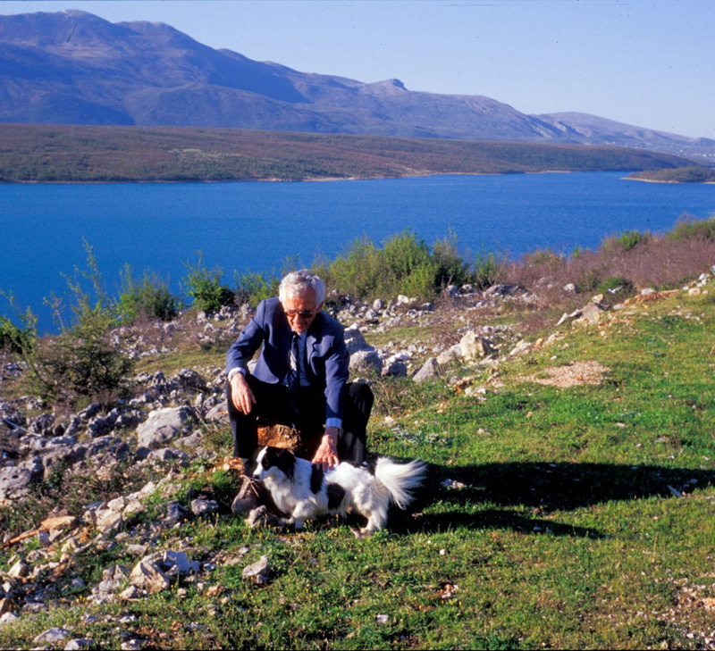
Early life and education I was born 70 years ago in the small town of Sinj, Dalmatia, Croatia, Yugoslavia. When I was 5 months old, my father was sent to prison for two years on Goli otok (Bare Island), an ill-famed prison for politically inappropriate people at that time. At 24 years old, he could not understand that Stalin, who was an idol for young Communists, had become an enemy overnight after the Informbiro Resolution, by which the Yugoslav Communist party was expelled from the international Communist party led by Stalin. It was a price for living free on the outer side of the Iron Curtain, between East and West, where Yugoslavia remained until the end of 1990, when the Balkan wars started. My father has stuck with Communist ideas until today, almost 95 years old (Figure 1). Under his influence, I entered the Communist party in high school, but very quickly realized that politics is not something I would like to practice.
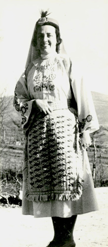
Figure 1. My father and our late dog Maša 15 years ago. After primary school, I attended Classical Gymnasium “Natko Nodilo” in Split, Croatia, where, in addition to other high school subjects, I learned Latin and Ancient Greek. At that time, my life consisted of studying, taking care of my two younger brothers, and helping my mother in everyday domestic jobs. After gymnasium in 1969, I decided to study medicine, mainly because it was not available in Split at that time. I wanted to distance myself from my primary family to escape my mother’s control. I wanted to be free. My mother supported me in my decision (Figure 2). I finished Faculty of Medicine at the University of Zagreb in January 1975. After one year of obligatory practice, my first employment was in the emergency clinic in Karlovac, where I worked for three years alternating between twelve-hour day and night shifts, as I traveled from my residence in Zagreb. It was a very stressful job for a young, inexperienced M.D., and I was terrified of the possibility of doing something wrong. I was looking for a residency in pathology in Zagreb, but the salary for a beginner was so small that I could not pay rent for my small apartment. I therefore moved to a small town in Slovenia, Ravne na Koroškem, which has a large iron factory and where I in cared for factory workers’ health. I received an apartment from the Health Centre, only having to pay a small rent.
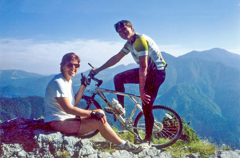
Figure 2. My mother in her youth in the national costume of continental Dalmatia, which she was wearing just for the photo. My family I met my husband Srđan during my third year of medical faculty. He had just entered the Faculty of Mechanical Engineering. He was a city boy from Zagreb, full of energy, honest and kind. He introduced another dimension into my life – fun and entertainment. Every weekend we spent together, we joined his friends and enjoyed life. Three years later, I completed my study of medicine. We married in 1978, just before he was conscripted into the army for one year.
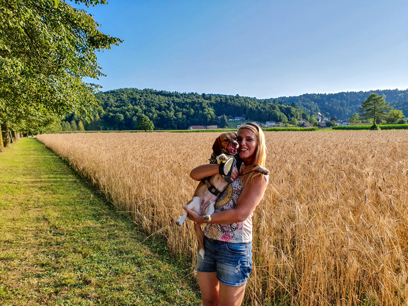
Figure 3. Me and my husband long ago on the top of one of the Slovene hills we had reached by bike. After military service of one year, my husband joined me in the small town in Slovenia, but he was not very happy there. He missed the greater opportunities of city life. Our daughter Saša was born in 1980, and our son Marko in 1983. In 1982 I obtained a residency in pathology in the nearby small town of Slovenj Gradec, and went to study pathology at the Institute of Pathology, Faculty of Medicine, University of Ljubljana (IP FM UL). After two years of residency, the Institute offered me a staff position, and I accepted. My husband was very happy. He obtained a good position at an engineering company in Ljubljana and we have remained in Ljubljana to this day. My husband has supported me throughout my career, taking care of our children, but asking that the weekends must be ours (at least Sundays). So throughout our life together we spent our weekends practicing many sports: tennis, jogging, hiking, mountain climbing, cycling. During the summer we would wind-surf as well, and during the winter we would ski (and later cross country skiing). We became serious mountain bikers, practicing twice a week for years (Figure 3). Last year, for my 69th birthday, I switched to an e-MTB bike. I use only eco power, the smallest of five levels of support, but it helps me a lot and I no longer suffer. Our daughter Saša is an architect, single, loves cats and especially loves her dog Cloe, a young beagle with a particularly stubborn character (Figure 4). Our son Marko is a happily married father of two sons (Figure 5).
Figure 4. Our daughter Saša and her dog Cloe.
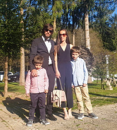
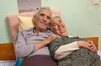
Figure 5. Our son Marko with family. My mother (91) and my father (almost 95) live in Split, in their own home, with no sign of dementia and without any medication except vitamin B12 for my mother, who looks after the two of them (Figure 6). My father goes to a nearby café every day to drink coffee and read the newspaper, and my mother goes every day to the market across the street to buy fresh vegetables, fish, fruit and other things. Last year, they celebrated 70 years of marriage.
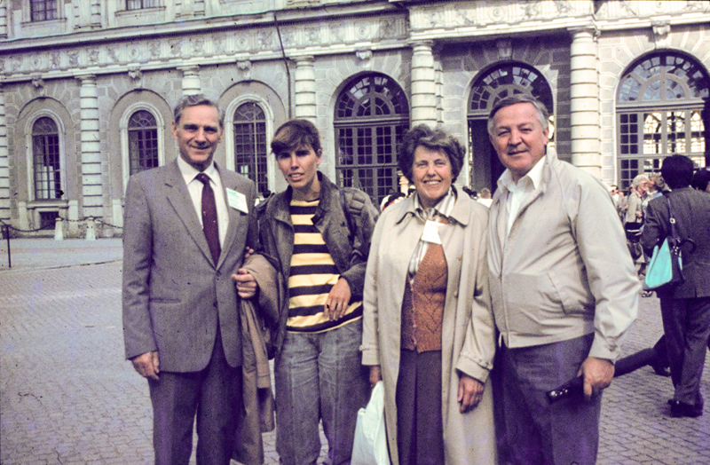
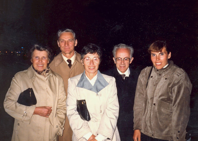
Figure 6. My parents, 94 and 90 years old, last year. Neuropathology In the third year of my residency in pathology, in 1984, Prof. Danilo Tavčar, the pathologist who established the department of neuropathology (NP) at the IP FM UL in 1972, invited me to join the department and become his assistant. I was also invited by Prof. Nina Gale, who was head of the department of head and neck pathology. I needed some days to decide which invitation to accept. Prof Tavčar was almost 70 years old, and I would very soon remain alone in a quite unknown field of pathology. Nina Gale was only a little older than me, a very successful and prominent head and neck pathologist. It would be much easier to accept her offer, but the unknown field of NP attracted and challenged me more. So I accepted Prof. Tavčar’s offer, and I did not regret the decision, even though it was not easy. I worked with Prof. Tavčar for two years parallel to my residency in pathology, which I completed in 1986. In the meantime, I attended postgraduate study for neurology in Zagreb, which lasted four months during the academic year 1984/1985. My working days were in Zagreb, and I spent the weekends with my family in Ljubljana. My husband took care of our 4-year-old daughter and our 1.5-year-old son spent the working days with my husband’s parents in Zagreb, and travelled with me for weekends to Ljubljana. Prof. Tavčar took me to the International Congress of Neuropathology in Stockholm in 1986 where we met Srečko Pogačar (1929 to 2018) (Figure 7), a Slovene neurologist and the first Slovenian neuropathologist to have gone to Rhode Island Medical Centre in the USA to study neuropathology, in 1965. Srečko had been the first to establish a NP lab at the Clinic of Neurology in Ljubljana in the early sixties. When he came back from the USA to Ljubljana, the NP lab no longer existed. Disappointed, Srečko decided to leave Slovenia and continue his career in the USA practicing psychiatry and neuropathology, especially brain cuttings. He was a lecturer on neuropathology and neurology at Harvard and Brown Universities [1].
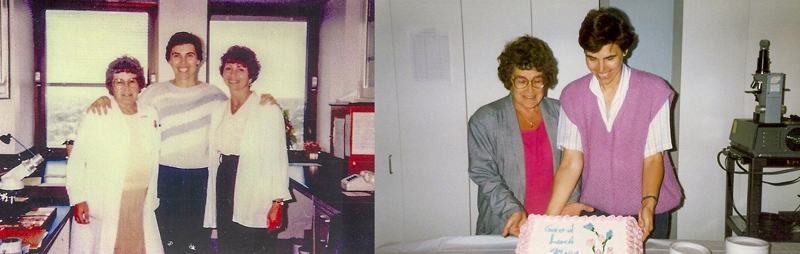
Figure 7. International Congress of Neuropathology Stockholm 1986. From the left: Prof. Danilo Tavčar, myself, Prof. Tavčar’s wife Maca, and Srečko Pogačar. Prof. Tavčar undertook further training in NP in Massachusetts General Hospital, Boston, under Dr. E.P. Richardson, for six months in 1974. He shared all his knowledge of NP with me. In the first half of the 1980s, immunohistochemistry started to become a useful tool in pathology. It was a very attractive challenge for me. Prof. Tavčar left me to do that job. After we returned from Stockholm, he experienced an epileptic attack in his left leg – Jackson’s epilepsy - for the first time. He knew what it meant. After a CT-scan, which showed a focal lesion in the right parietal lobe, on which he was operated, he did not want to see the biopsy. He worked until spring 1987, going to radiotherapy from his office, and did not want to take any of the alternative therapies that his wife had asked him to. He died in December 1987, twelve months after his first epileptic attack. I stayed in contact with Srečko Pogačar, who offered to organize a six-month fellowship for me at Rhode Island Hospital (RIH) in Providence, where I could learn neuropathology under the mentorship of the now deceased Dr. Mary Ambler, a neurologist and neuropathologist. It was a great opportunity that I had to seize. I reached an agreement with the current chief of IP MF UL, prof. Dušan Ferluga, and with my family. My daughter was 9-years-old and my son 6-years-old. My husband had a good job in Ljubljana and could not drop everything to go with me. I was enthusiastic and happy for the opportunity to be more confident in NP. I remember the early morning in Zagreb when my husband drove me to the airport. My children stayed with their grandpa and grandma. I had a cramp in my epigastrium from the time I boarded the airplane in Zagreb until I landed six months later at the same airport and hugged my children and my husband. At that moment the cramp disappeared. Professionally and otherwise, I had a good time in the USA, but I missed my family a lot. No Skype, no Facebook, no SMS, no WhatsApp, no e-mail. Only Atlantic surface mail and expensive telephone calls once a week with my husband.
Figure 8. Six months training in neuropathology in Rhode Island Hospital. A. Mentor Mary Ambler, me and Rosemarie Guglielmi. B. Mary Ambler organized a leaving party for me. Dr. Mary Ambler was a great person and teacher, and her technician Rosemarie Guglielmi still remains my dear friend (Figure 8). While in the USA, I spent one week in the Department of Pathology at Cleveland University Hospital, where I worked with another Slovene, Uroš Roessmann, whom I met at ICNP in Stockholm in 1986 (Figure 9). He gave me the opportunity to see more child brain tumours, which RIH did not have. Uroš became a good friend and, after he retired in 1991, he visited Slovenia twice, spending three months each time at the IP MF UL as a visiting professor. It was another valuable opportunity to learn more from an experienced neuropathologist.
Figure 9. International Congress of Neuropathology Stockholm 1986. From the left: Maca and Danilo Tavčar, MIlči and Uroš Roesmann, and me. Before going to the USA, I had joined the research group of Janez Sketelj (Figure 10), pathophysiologist, eminent researcher and head of the Institute of Pathophysiology MF, UL. We had planned research on the regeneration of the rat sciatic nerve after various types of injury, which I continued to pursue after coming back to Ljubljana. This research resulted in my MSc in 1992, PhD in 1996 and four articles [2-5].
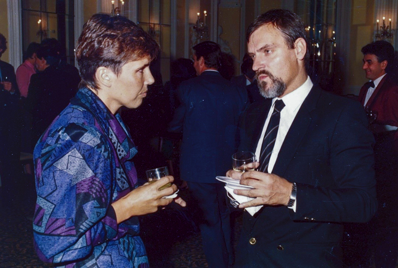
Figure 10. Janez Sketelj, my mentor for MSc and PhD. I was left without a mentor or colleague at the Institute who could help me in cases of rare brain tumours and brain pathology. In September 1987, I attended the Danube Neurology Symposium in Innsbruck, where I met Herbert Budka, a well-known neuropathologist and head of the Institute of Neurology in Vienna (Figure 11).
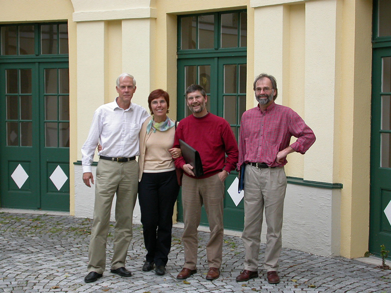
Figure 11. Herbert Budka, who I met for the first time at the Danube Symposium of Neurology in Innsbruck in 1987. Throughout almost my entire career in NP, until his retirement in 2011, he and his staff were a great help to me and to Slovene NP. I visited them several times and spent three months with them in 2007. It was an excellent way of learning, through a daily microscopic session with Herbert and all the others around a multiple head microscope. I remember asking Herbert why they had to look at every meningioma. “You have to see a lot of ordinary meningiomas to recognize a peculiar one” Herbert said, and he was right. In 1995, Herbert Budka invited Slovenia to join a CJD surveillance project led by him and financed by the EU. I have very pleasant memories of the annual meetings in Baden near Vienna, with excellent scientific and social programs, where I learned a lot about prion diseases and had an enjoyable time with Scandinavian neuropathologists (Figure 12). As a result of that program, IP MF UL established a Slovene CJD surveillance system together with the National Institute of Public Health and Slovenian neurologists [6, 7]. IP MF UL became the referral centre for CJD in Slovenia. To date, we have confirmed 88 sCJD cases, one genetic CJD, two cases of Gerstmann-Sträussler-Scheinker syndrome (GSS, a a mother in 2007 [8] and her son in 2020). A case of clinically obvious fatal insomnia with a new N181S PRNP mutation, without family history, was recognized in 2015. Despite regulations surrounding this project by Slovenian law in 2001, some clinically suspected CJD cases and the case of fatal insomnia escaped autopsy. Fifty-three clinically suspected CJD cases were disproved by autopsy. A Prion laboratory with containment level 3 was established in 2000, supported by the Ministry of Health and stimulated by the work of Vladka Čurin Šerbec and her team from the Centre for Transfusion Medicine in Ljubljana, who produced an anti-PrP antibody named V5B2 with the same quality as other commercially available anti-PrP antibodies. Four PhDs and several valuable papers have been produced in that lab [9-12]. Thanks to Herbert and the CJD surveillance system, the annual incidence of sCJD increased in Slovenia from 0,5 per 1 mil (in the period 1985-2000) to 1,5 per 1 mil (in the period 2001-2020). In the last ten years, specifically, the incidence reached 2,5 per 1 mil [13, 14]. Slovenia is still a member of the CJD surveillance project, which is now lead by the United Kingdom. We have not identified any variant CJD (vCJD) cases in Slovenia, however a case of sCJD, MV2A molecular subtype, had clinical, radiological and pathological similarities with vCJD [15].
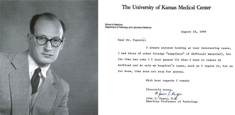
Figure 12. Scandinavian neuropathologists, from the left: Sverre Mork, me, Peter Stubbe Teglbjaerg and Henning Laursen. Excellent company at the annual CJD meetings in Baden by Vienna. My collaboration with the Institute of Neurology at AKH in Vienna was also fruitful in tumour pathology. Herbert Budka, Christine Haberler, Johannes Hainfellner and Ellen Gelpi were of great help in my rare and difficult brain tumour cases, which resulted in several papers [16-18]. Ellen Gelpi and Gabor G Kovacs recognized one of our cases of brainstem tauopathy [19], to probably be a new entity of anti-IgLON5-related tauopathy [20].
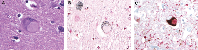
Figure 13. A. The late John Kepes, an excellent neuropathologist and person. B. His letter announcing that he would no longer be available for consultation. Another neuropathologist from the USA also provided me with great help, the late John Kepes, who was a very nice person. Not only would he send me his opinions on cases I had sent him for consult, but he also sent microphotographs to point out the main changes important for the case diagnosis. I was very touched when he sent me a letter with an apology that he was unable to help any more (Figure 13). I developed my academic career at the IP MF UL and became full professor of pathology in 2011. Even though it is my duty to research, I am not a researcher at heart. I don’t like statistics but I really enjoy studying every brain, especially neurodegenerative ones. During my career I have attended all European and some International Congresses of NP, presenting these cases (my CV in Supplement). I would like to highlight some of which I am proud. I identified and published neuropathological changes in four family members with a P364S MAPT mutation. The mutation was very new and had been described by Italian researchers two years previously [21]. They described the clinical presentation of the patient, made recombinant tau with the mutation, and showed that under in vitro conditions “P364S tau revealed a remarkable increase in aggregation rate with respect to WT tau, displaying mostly protofibrils and some short fibrils after 24 hours, and a consistent presence of both straight and twisted long fibrils after 5 days. The propensity of P364S tau to aggregate was even higher than P301L tau used as well-known mutated control.”
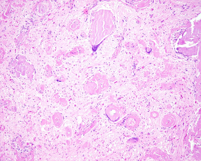
Figure 14. Composite neuronal tau inclusion (CNTI) in a family with a P136S mutation in MAPT. A. H&E, B. Gallyas silver staining displays only the peripheral part of the inclusion, which is 4RTau positive, while the central part is only 3RTau positive in double immunohistochemistry on 3R and 4R Tau (in C). I recognized that this mutant tau produced in all affected family members a new neuronal tau inclusion consisting of two components (Figure 14). We designated it Composite Neuronal Tau Inclusion (CNTI) [22]. Additionally, I discovered that in one family member’s brain, in addition to CNTI, all other neuronal tau inclusions known so far were present [23]. Another interesting result of brain research was a case of a young male with paranoid schizophrenia, who developed bulbar symptoms and died in his sleep. A few months before death, he was diagnosed by MRI as having multiple sclerosis. The brain cutting revealed that he had light chain deposition disease (LCDD) brain vasculopathy with multifocal hypoxic brain injury, especially prominent and fatal in the medulla (Figure 15). When we submitted the manuscript to the journal Human Pathology, it was the first case of LCDD restricted to the brain [24]. By the time of publication, however, the first published case of LCDD restricted to the brain had been published by Fischer et al. [25]
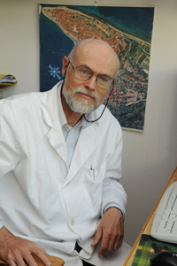
Figure 15. Severe light chain deposition disease vasculopathy (LCDDV) of medulla. H&E, original magnitude 40x. My most exciting and successful research in brain pathology was a case of Zika virus-induced microcephaly, with the first confirmation of causality displaying the virus in the foetal brain by PCR, immunohistochemistry using maternal sera, and by electron microscopy [26]. Several papers were produced on this case thanks to our young resident at that time, Jernej Mlakar, who performed a perfect brain autopsy (Figure 16) [27-29]. Jernej is now a general pathologist involved in NP together with three other fields of pathology. I was lucky and privileged only having done NP throughout my career as a pathologist. Even though I was a solitary neuropathologist for most of my NP career, several valuable people were included in NP, without whom my work would not have been possible. Two excellent NP technicians, Dori Jazbec and Marija Zupančič; two administrators, Marija Blejc and Mojca Keber; immunohistochemical masters, Daniel Velkavrh, Majda Dimnik, Miša Omerzel and Ajla Hajrlanović; geneticists Alenka Matjašič, Ema Boštjančič and Andrej Zupan (the most invaluable people in degenerative and especially in tumour brain pathology today) [8, 15, 22, 23, 30-32], and brain cutting assistants, Janez Caserman, Srđan Čekić and Damir Novak. Last but not least, I should mention the numerous students and residents of pathology, neurology and neurosurgery to whom I have tried to impart all my knowledge of NP. I have also learned a lot from them. Many of the residents prepared clinico-pathological conferences, which we had almost every fourth Wednesday of the month before the coronavirus pandemic.
Figure 16. Me (actual) and Jernej Mlakar, future head of Department of Neuropathology at the Institute of Pathology, Faculty of Medicine, University of Ljubljana, Slovenia. So I am approaching the end of my almost 37-year career in NP. I have never regretted my choice, and even though I was the only neuropathologist in Slovenia most of the time, I have always favoured Mondays to Fridays. I know that after the 1st July 2021, when my retirement will start, my life will not be the same. I will miss my beloved NP a lot. Acknowledgment I am deeply grateful for technical support to our IT team, Metod Perme and Miha Juvan and our researcher, Nina Hauptman, who have always been available when my PC did not obey. New technology is great and very useful, but never makes mistakes, which is unbearable. References 1. Pogacar, S. and M. Popovic, Dr. Srecko Pogacar: from a castle in Slovenia to a clinic in RI. R I Med J (2013), 2013. 96(10): p. 38-40. 2. Popović, M., M. Bresjanac, and J. Sketelj, Regenerating axons enhance differentiation of perineurial-like cells involved in minifascicle formation in the injured peripheral nerve. J Neuropathol Exp Neurol, 1994. 53(6): p. 590-7. 3. Sketelj, J., M. Bresjanac, and M. Popović, Rapid growth of regenerating axons across the segments of sciatic nerve devoid of Schwann cells. J Neurosci Res, 1989. 24(2): p. 153-62. 4. Popović, M., M. Bresjanac, and J. Sketelj, Role of axon-deprived Schwann cells in perineurial regeneration in the rat sciatic nerve. Neuropathol Appl Neurobiol, 2000. 26(3): p. 221-31. 5. Popović, M., J. Sketelj, and M. Bresjanac, Changes of Schwann cell antigenic profile after peripheral nerve injury. Pflugers Arch, 1996. 431(6 Suppl 2): p. R287-8. 6. Popović, M., et al., Creutzfeldt-Jakob disease in Slovenia from 1985 to 2003. Wien Klin Wochenschr, 2004. 116(15-16): p. 524-9. 7. Budka, H., Concern about mad cow disease: end of the beginning, or beginning of the end? Wien Klin Wochenschr, 2004. 116(15-16): p. 505-7. 8. Kojović, M., et al., De novo P102L mutation in a patient with Gerstmann-Sträussler-Scheinker disease. Eur J Neurol, 2011. 18(12): p. e152-3. 9. Curin Serbec, V., et al., Monoclonal antibody against a peptide of human prion protein discriminates between Creutzfeldt-Jacob’s disease-affected and normal brain tissue. J Biol Chem, 2004. 279(5): p. 3694-8. 10. Colja Venturini, A., et al., Anti-idiotypic antibodies: a new approach in prion research. BMC Immunol, 2009. 10: p. 16. 11. Lukan, A., et al., Regional distribution of anchorless prion protein, PrP226*, in the human brain. Prion, 2014. 8(2): p. 203-9. 12. Vranac, T., et al., A single prion protein peptide can elicit a panel of isoform specific monoclonal antibodies. Peptides, 2006. 27(11): p. 2695-705. 13. Čakš Jager, N., et al., Analysis of 22 Years of Surveillance for Prion Diseases in Slovenia, 1996 to 2017. Zdr Varst, 2018. 57(4): p. 227-33. 14. Rus, T., et al., High Incidence of Sporadic Creutzfeldt-Jakob Disease in Slovenia in 2015: A Case Series. Dement Geriatr Cogn Dis Extra, 2018. 8(1): p. 42-50. 15. Bošnjak, M., et al., A case of MV2K subtype of sporadic Creutzfeldt-Jakob disease with florid-like plaques: Similarities and differences to variant Creutzfeldt-Jakob disease. Neuropathology, Published online April 5, 2020. 16. Gelpi, E., et al., Pleomorphic xanthoastrocytoma with anaplastic features presenting without GFAP immunoreactivity: implications for differential diagnosis. Neuropathology, 2005. 25(3): p. 241-6. 17. Filbin, M.G., et al., Developmental and oncogenic programs in H3K27M gliomas dissected by single-cell RNA-seq. Science, 2018. 360(6386): p. 331-35. 18. Clarke, M., et al., Infant High-Grade Gliomas Comprise Multiple Subgroups Characterized by Novel Targetable Gene Fusions and Favorable Outcomes. Cancer Discov, 2020. 10(7): p. 942-63. 19. Pretnar-Oblak, J., et al., Isolated bulbar paralysis in a patient with medullar tau pathology: a case report. J Neurol Neurosurg Psychiatry, 2010. 81(8): p. 847-9. 20. Gelpi, E., et al., Neuropathological criteria of anti-IgLON5-related tauopathy. Acta Neuropathol, 2016. 132(4): p. 531-43. 21. Rossi, G., et al., New mutations in MAPT gene causing frontotemporal lobar degeneration: biochemical and structural characterization. Neurobiol Aging, 2012. 33(4): p. 834.e1-6. 22. Popović, M., et al., Tau protein mutation P364S in two sisters: clinical course and neuropathology with emphasis on new, composite neuronal tau inclusions. Acta Neuropathol, 2014. 128(1): p. 155-7. 23. Štrafela, P., et al., Familial tauopathy with P364S MAPT mutation: clinical course, neuropathology and ultrastructure of neuronal tau inclusions. Neuropathol Appl Neurobiol, 2018. 44(6): p. 550-62. 24. Popovic, M., et al., Light chain deposition disease restricted to the brain: The first case report. Hum Pathol, 2007. 38(1): p. 179-84. 25. Fischer, L., et al., A 19-year-old male with generalized seizures, unconsciousness and a deviation of gaze. Brain Pathol, 2006. 16(2): p. 185-6, 187. 26. Mlakar, J., et al., Zika Virus Associated with Microcephaly. N Engl J Med, 2016. 374(10): p. 951-8. 27. Onorati, M., et al., Zika Virus Disrupts Phospho-TBK1 Localization and Mitosis in Human Neuroepithelial Stem Cells and Radial Glia. Cell Rep, 2016. 16(10): p. 2576-592. 28. Štrafela, P., et al., Zika Virus-Associated Micrencephaly: A Thorough Description of Neuropathologic Findings in the Fetal Central Nervous System. Arch Pathol Lab Med, 2017. 141(1): p. 73-81. 29. Vesnaver, T.V., et al., Zika virus associated microcephaly/micrencephaly-fetal brain imaging in comparison with neuropathology. Bjog, 2017. 124(3): p. 521-25. 30. Matjašič, A., et al., A novel PTPRZ1-ETV1 fusion in gliomas. Brain Pathol, 2020. 30(2): p. 226-34. 31. Matjasic, A., et al., Expression of LOC285758, a Potential Long Non-coding Biomarker, is Methylation-dependent and Correlates with Glioma Malignancy Grade. Radiol Oncol, 2017. 51(3): p. 331-41. 32. Matos, B., et al., Dynamic expression of 11 miRNAs in 83 consecutive primary and corresponding recurrent glioblastoma: correlation to treatment, time to recurrence, overall survival and MGMT methylation status. Radiol Oncol, 2018. 52(4): p. 422-32.
Copyright: © 2020 The author(s). This is an open access article distributed under the terms of the Creative Commons Attribution 4.0 International License (https://creativecommons.org/licenses/by/4.0/), which permits unrestricted use, distribution, and reproduction in any medium, provided the original author and source are credited, a link to the Creative Commons license is provided, and any changes are indicated. The Creative Commons Public Domain Dedication waiver (https://creativecommons.org/publicdomain/zero/1.0/) applies to the data made available in this article, unless otherwise stated. |