|
|
|
Free Neuropathology 1:29 (2020) |
|
Reflections |
|
Neuropathology as a Life-Task |
|
Werner Jänisch |
|
Address for correspondence: |
|
Submitted: 12 September 2020 Accepted: 13 October 2020 Copyedited by: Henry Robbert and Biswarathan Ramani Published: 16 October 2020 |
|
Additional resources and electronic supplementary material: supplementary material |
|
Keywords: Neuropathology, Personal reflections, GDR, Charité |
|
1. Prologue Neuropathology as a special field in medicine saw its development not until the second half of the twentieth century. Its roots are in neurology and pathology. Its significance has risen because of the emergence of neurosurgery. The validity of neuropathological diagnoses greatly increased especially because of the application of immunohistochemistry and genetics. This may have been the rationale for the editor of this journal to have neuropathologists’ paths of life delineated for future generations under the heading “Reflections.” This includes the various pathways and detours to neuropathology and the ways research findings have been generated, which may eventually prove important or useless. It has been the desire of the editor that authors not only reflect on their professional activities and experiences, but also present themselves as persons. This paper should not be intended as an autobiography, but the development and experiences of the author be presented in appropriate length. I think of this as a good approach, which I will attempt to pursue. 2. Social origin and education I was born in 1932 in the German Reich, specifically in the industrial city of Chemnitz, Saxony. My parents married in 1928 and lost employment shortly after due to a hard-hitting economic crisis. Even as my father successfully found work as a truck driver the income of my parents was so low and the living conditions so confined that I remained their only child. My childhood was overshadowed by the reign of National Socialism in Germany. In 1938, I entered the Volksschule (i.e., the prevalent type of elementary school at that time). In the beginning, I felt enthusiastic about a young, skilled teacher. When he did not return after the summer break of the year 1939, I and the other kids were very disappointed. The teacher had been drafted as a soldier by the Wehrmacht. He was replaced by an old teacher who approached school lessons by using a cane for whipping and punching, thus taking away our joy in studying. In the Volksschule we learned how to read, write, basic math and Adolf Hitler’s curriculum vitae. Many classes were replaced by foot drills in the schoolyard. They were directed by a teacher who had suffered a brain injury in the beginning of the war, disqualifying him from military service. By the end of 1943, when I, a mediocre student, had reached sixth grade, classes were suspended in the larger cities of the German Reich. This was due to the alleged danger posed by Anglo-American air strikes. My father was drafted as an ordinary soldier at the beginning of the war in 1939, finally became an Unteroffizier (i.e., roughly on the level of a sergeant) and died as a prisoner of war in a military hospital in September of 1946. My hometown Chemnitz was spared from air strikes until the beginning of 1945, but was largely destroyed on March 5, 1945 by Anglo-American bomber squadrons. Our two-room apartment, which was located in a big apartment complex, was affected by that, as well. My mother and I survived, became homeless and possessed only what we had on our bodies. We were placed in an emergency shelter in the small town of Mittweida, 18 km north of Chemnitz. In mid-April of 1945, this area was occupied by American troops without a fight. These troops left after about two weeks and a few days later troops of the Red Army took over the town, because it was part of the designated Soviet occupation zone. A Soviet military administration had control over this zone. One of its first actions was the order to vaccinate the entire population. This was a life-saving measure because of the aftermath of the war and the refugee streams infectious diseases such as typhus, diphtheria and tuberculosis were spreading out quickly. For me personally there was another order of the Soviet military administration that was of great importance: It was said that beginning on October 15, 1945, classes were to resume. This meant that I, because of my age at that point, was able to get into eighth grade of the Volksschule. Together with 44 other boys, I joined class led by a teacher who was an elevator installer but had been prepared for teaching in a crash course. My knowledge gaps were vast, which was also the case with other students who went through similar experiences as I had. My teacher tried to make up for this with remarkable patience and a lot of empathy. However, it should be said that it helped that most students were very eager to study. Our thirst for knowledge was insatiable. I am grateful to this teacher for providing us with a solid base of knowledge and teaching us important basic principles in ethics. Unfortunately, eighth grade was the last grade of the Volksschule. Graduates were instructed in a profession, or they entered professional life without any instruction. As a child, I wanted to become engine driver of a steam locomotive. The requirement for this was the professional instruction in a railroad facility. A few weeks before graduation, I applied for such an apprenticeship and got a rejection. Deeply disappointed, I started looking for an alternative. Mittweida was part of the textile industry. Workers were able to work in spinning mills, weaving mills and sewing rooms without instruction (unlike their superiors). These places also offered no apprenticeships. The only choices I had were to become instructed as a painter, carpenter, metal worker, chimney sweeper, baker or butcher. None of these prospects was desirable. In this situation suddenly a new perspective opened up due to another order of the Soviet military administration: It was said that skilled graduates of the Volksschule were able to enter the Oberschule (roughly equal to high school). Until then parents were only granted to put their children into Oberschule (which are called Gymnasium nowadays) after they had finished fourth grade – but only in case they were able to pay tuition. This was not the case for me. My teacher made arrangements so that I could continue my studies without having to pay tuition! My mother and I were thrilled. I initially joined a class for students who had to adapt, which means that lessons were conceptualized as to make sure students could gain the knowledge the other students had been building on since fifth grade of the Oberschule. I was an enthusiastic student. Latin, biology, chemistry, history, German literature and Russian became my favorite subjects. Math was not one of those, although we had a good teacher. I also encountered problems in English. I was not aware that English words were pronounced differently as could be assumed from the way they were written (for example, it was not clear to me how to pronounce the “u” in a word like “must” the way it sounds in English as opposed to a “u” in German). The English teacher, a former Protestant minister, never imparted this knowledge to us. Hence, I eagerly studied the vocabulary from the book but still had bad grades. After a couple of months the teacher left the school, so English lessons were suspended because there were not many English teachers left in the Soviet occupation zone. Because unemployment was widespread in the other occupation zones in the years 1946–1948, workers came to the Soviet occupation zone, including English teachers. One of them came to our school and with him his unconventional methods. We have been studying Walter Scott’s novel “Ivanhoe” for a full school year. He distributed copies of the novel’s German translation, told us to read it chapter by chapter, and demanded summaries and interpretations of its content in German. This became the basis for our grades. This way I was able to study an important work of English literature and got good grades. However, I did not learn a single word of English and I was aware of this. I became friend with a student of a higher grade who had English classes prior to 1945. He agreed to teach me some of his knowledge. This way I was able to improve my pronunciation of the vocabulary and learn some basic grammar. But still my English skills remained utterly mediocre. I was able to read and understand simple texts, but I was lacking an active command of the language. Because I took great joy in studying, my time in the Oberschule went by in a flash. A few months before the Abitur (i.e., the high school diploma), we were asked to decide if we planned to continue studying, which subject we wanted to major in, and at which university we would try to apply. Since I had been an eager student of biology in school and extended my knowledge reading zoological books and spending time in nature, I knew early that I wanted to study biology. But suddenly doubts arose: What would I do if biology would not meet my expectations, if chemistry and physics were more interesting to me? This led to the idea of studying medicine. It includes morphological subjects, microbiology, genetics, biochemistry, pharmacology, applied radiophysics and I could choose which scientific area I wanted to pursue later in my career! The next question was concerning the place I wanted to study. Then, in the GDR (German Democratic Republic, DDR), there were medical schools at the universities in Berlin, Leipzig, Jena, Rostock and Greifswald. Because of my interest in biology I was leaning towards Jena early on, even though I had never visited the city. The reputation of the biologist Ernst Haeckel and the anatomist Christian Loder made that university so attractive to me, which was why I applied to this school. Since I graduated from the Oberschule with the final grade “sehr gut” (i.e., the best overall grade) in 1951, I was promptly enrolled as a student of medicine and received a monthly stipend of 260 Mark. It should be noted that in the GDR every student could apply for a scholarship, which was not a loan and did not have to be paid back later. The sum of the stipend was dependent on the final grade in high school and the grades a student got at the university and could vary between 180 and 260 Mark. The sum of the stipend I received was sizable for me personally (my mother, who was full-time employed, earned much less than that). It should be noted that I paid my landlady 20 Mark per month, that 1 kg of bread cost 30 Pfennige (pennies) and one meal in the dining hall 70 Pfennige. After my first year I completed my exams in physics, zoology and general botany with the best grade (“sehr gut”) and was ordered to the Dean’s office. I was offered the possibility to continue my studies at a university in the Soviet Union. I was selected because of my academic performance so far, and my high school grade in Russian. I asked for some time to think because I wanted my mother to agree. This was necessary, because she would be by herself for a long time if I went to study abroad. Studying in Jena, I was able to visit her for a weekend every two months. Without any hesitation she recommended me to take the offer. So in October of 1952 I went to Moscow with about 200 other students of various disciplines and from there went to Leningrad to study medicine. There were three major civil medical schools with different instructional foci and a military medical school. The different foci of the schools can be explained by the fact that most graduates are sent to different parts of the country without receiving a specialist medical instruction. In remote places of the country (Siberia, north of the polar circle, etc.) it was not uncommon for them to be the solely responsible physician. There was no telemedicine back then. I arrived at a medical school for future pediatricians. Today this is the Saint Petersburg Academy of Pediatrics and Maternity. Its students came from all republics of the Soviet Union and many foreign countries. All special areas of medicine were taught, as was the case in German universities as well. What was special about this medical school was that there was a special focus on children. This concerned infectious diseases, nutritional disorders, and skin diseases, among others. But this also concerned theoretical subjects such as physiology, pathology (pediatric pathology was taught very extensively) and even forensic medicine. The instruction of the students took top priority, was very thorough and frequently individualized. Interested students were offered “academic student circles” in different special areas and could thus learn about and participate in professors’ research activities. I took part in this as well, first in anatomy and later in pathology. In 1958, my studies concluded with a state exam “with distinction” (“Ausgezeichnet”), and I returned to the GDR.
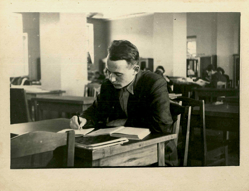
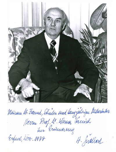
Fig. 1: In the reading hall of the dormitory in 1953. 3. Memories of Leningrad After arriving in Leningrad, I was lodged in a dorm room. The dormitory’s construction began before World War II and was finished after the end of the war, partly with the help of German prisoners of war. It was clean and cozy. On the ground floor there was a library with a reading room offering literature relevant for studies and the belles lettres that could be borrowed and studied (Fig. 1). All newly arrived foreign students could take part in a tour of the city. Since the city underwent restorations shortly before, the historical buildings were shiny and the parks well maintained. We were shown the memorials of Tsar Peter the Great and Tsar Catherine the Great and various cultural sites. I used to go to the theater frequently to enjoy the works of Pushkin and Chekhov in the beauty of their language, to opera performances and the ballet of Leningrad, and occasionally, symphonies, as well. The latter was directed by Kurt Sanderling, who during the reign of National Socialism emigrated to the Soviet Union and later acted as chief conductor in the GDR. Leningrad presented itself as a vibrant big city with a dense net of buses, trolleybuses, and streetcars. Back then underground railways did not exist yet. As a German student I was warmly welcomed and offered help by the university and the general population. Never was I or, as far as I know, any other students from the GDR blamed for the drastic crimes that happened during the German blockade of Leningrad. We received a monthly stipend of 800 rubles. (Later the ruble, and the stipends, was devalued 10 to 1.) The dorm room cost 15 rubles per month. It should be noted that the monthly salary of our professors were between 1,200 and 1,600 rubles. Hence, we could dedicate ourselves to our studies without having any material worries. My fellow students welcomed me with friendliness. They were eager to get to know how we were living in Germany and how we experienced the war. The first question I was asked was: “Is Hitler still alive?” Because of my imperfect command of the Russian language, I thought I misunderstood that question, because for us Germans it was clear without a doubt that Hitler was dead. Until some back and forth it became clear to me that even in 1952 the people still worried that Hitler might have escaped and could become dangerous again. I asked many questions, too, and got to know a lot about the reality of everyday life and life during the war. Successively I learned to understand the Russian people’s way of thinking. One example from the first days of my stay might make this clear: I asked a Russian student, whom I shared a dorm room with the question: “Valentin, where is it you are from?” He replied: “prigorod”, which translates to suburb, i.e. close to Leningrad. Later I found out that the distance between the two places was 800 km. I got further insight when I was invited to the homes of university staff. I was introduced to Russian hospitality and their difficult living conditions. One of the families, which consisted of two parents and two small daughters, lived in a room of about 40 m² in what was called “communal quarter”. These were large apartments consisting of eight or nine rooms in houses spanning four stories constructed during the reign of the Tsar. Because of the scarcity of housing, these large apartments were divided and rented roomwise. The former kitchen that was accessible to all families was equipped with a big boiling pot for everyone and a small gas stove for each family. At the apartment’s entrance, there was a doorbell and a list with the family names of the renters accompanied by a number of ringtones. My hosts had the number seven. When the ringtone occurred, somebody in the family answered the door. During my stay with them and during intense conversations the doorbell rang frequently. When I said, “This is for you” they replied, calmly, “no, it was only six times” and continued talking to each other. I mention this because this situation was unusual to me and gloomy, but they showed to me that there are more important things, namely practicing hospitality, which they did extensively. This had an influence on me, so for my wife and me it was clear that we were hospitable to visiting students and fellow researchers, regardless from which country they came. I also feel deep gratitude for my professors and their assistants. They trained me how to do my first pathological/anatomical dissections. They introduced me to the great public library of Leningrad and showed me how to navigate the scientific catalogs. With their support I learned how to make microscopic preparations and use for my first academic publications. Saying farewell to Leningrad was hard for me. 4. Becoming a pathologist and my journey to neuropathology In medical school I was especially interested in morphological subjects: First anatomy, then pathology. As a student I conducted more than 100 autopsies by myself, as well as writing the dissection reports and participating in clinicopathological conferences. This was an occasion for me to delve deep into scientific literature. For example, performing a postmortem of a patient with an oligodendroglioma motivated me to buy and study K. J. Zülch’s book on brain tumors (23) in 1957. Back then, I was already intent on becoming active as a physician in the field of pathological anatomy. It was my desire to get instructed in this field at a university institute. In order to be granted a full medical license in Germany, one had to have practiced as an assistant in a clinical subject for at least one year. Therefore, in 1958 I applied to the Medical Academy Erfurt to be instructed for six months each in the clinics for internal medicine and surgery. In the clinic for surgery I was working in the department of neurosurgery, which was headed by Professor Usbeck. The department mostly treated patients with brain tumors and trigeminal neuralgia. At the end of this training Professor Usbeck offered me to train me as a neurosurgeon. I declined by explaining that I had decided to receive my specialized training at the Institute for Pathological Anatomy of the Medical Academy Erfurt. To this, he responded: “Then go ahead and take care of histopathological diagnostics of glioma. Almost every diagnosis I receive from the pathologists is astrocytoma. This cannot be right, because since Baily and Cushing (1) we know more about glioma and need exact diagnoses for our treatment strategies.” I took this suggestion seriously. First, because I knew from my own experience that at most German facilities for pathological anatomy – regardless if university or municipal – neuropathological questions did not get much attention. It was mostly neurologists and psychiatrists who were dealing with neuropathology. Secondly, I had realized that the field of pathology was so extensive and diverse that it was not possible to gain profound knowledge in every subfield. Furthermore, I desired to do research. Therefore I decided to pay special attention to the area of neuropathology during my training in pathology. This began with in-depth studies of the literature. The Institute for Pathology at the Medical Academy Erfurt, founded in 1954, had a well-equipped special library. Also, the academy had a central library, which allowed to order any missing scientific literature from all major libraries in the country and abroad for free. Thus, there was nothing in the way of further specialization. However, I needed Professor Harry Güthert to agree to my endeavors (Fig. 2). He became prosector of the Municipal Clinic in Erfurt in 1946. He became the director of the newly built Institute for Pathology when the Medical Academy Erfurt was founded in 1954. The institute also had a large barn for test animals. The institute’s staff comprised about 40 people, including two chief physicians (Oberärzte) and nine assistant physicians (Assistenzärzte). About six months after starting this activity I suggested to the director that I wanted to specialize in neuropathology. After a couple of minutes of thinking he replied: “Agreed. I will send at the University of Leipzig for a couple of weeks. There you can become familiar with the basics of neuropathology and study histopathological preparations.” He had his secretary immediately connect him to the director of that institute to arrange a date for my visit. I used to see again and again that this kind of rapid and focused reaction was typical of Professor Güthert’s style of leadership: When something was suggested to him that furthered the development of the staff and the institute he got behind those suggestions with all means that were at his disposal.
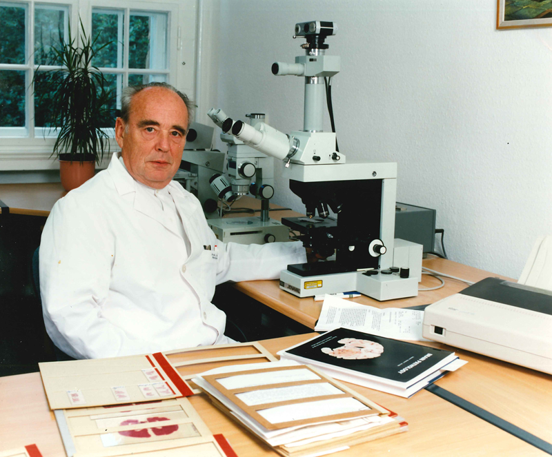
Fig. 2: Professor Harry Güthert, director of the Institute of Pathological Anatomy of the Medical Academy Erfurt in the year of his retirement. After returning from this stay I suggested to open a neuropathological laboratory at the Institute for Pathology in Erfurt. In Leipzig I was able to get to know the advantages of microscopic large sections for brain research and the meticulous photographic documentation of medical findings. Therefore, I asked Professor Güthert to acquire a microtome for large sections. Back then in Germany, the only producer of such devices was the company Jung in Heidelberg. The devices and the necessary equipment for preparing paraffin sections were rather expensive. Since the universities in the GDR were wholly financed by the state, the necessary foreign currency (West-German Deutsche Mark) had to be provided by the ministry of education. This did not pose a major challenge for the director, either. In the fall of 1960, he sent me to Heidelberg to the company Jung so I could select and order a device tailored to our needs. Furthermore, he arranged for me to get to know how to use the device at the Institute of Neuropathology at the University of Gießen during the same trip. He also granted me the support of a photographer (who was on the institute’s staff) whenever I needed informative macro shots of neuropathological findings to be produced with a plate camera. Hence, already at the beginning of my neuropathological activities, scientifically valuable documentations of medical findings could be generated in black and white, which I could later use for publications in books and journals. Back then, many diseases of the central nervous system (e.g., tumors, aneurysms, abscesses) were not surgically operated, because the diagnostics and, in part, surgical procedures had not been developed well enough (there were no special imaging techniques except for pneumencephalography and arteriography, and no microsurgical methods). Hence, at that time pathological findings of the brain without iatrogenic changes were not rare. Furthermore, in the GDR there was an obligation to autopsy people who died with suspected or evidenced malignant tumors, infectious diseases, stillborn and babies, people who died without explainable cause of death, suspected unnatural death, or when physicians had a scientific interest in the autopsy findings. Hence, autopsies could be conducted relatively often. For example, the staff of the Institute for Pathology at the Medical Academy Erfurt conducted between 2,000 and 3,000 autopsies annually. The share of autopsies with neuropathological findings was accordingly large. I did not conduct every single of these neuropathologically relevant autopsies; however, the decision to photographically document the findings and the diagnostic treatment of neuropathological findings was reserved for me. This way, after a very short period of time I had a relatively large, diverse wealth of material at my disposal, which could be used for in-depth scientific analysis and the training of students. I was involved in the latter fairly early, first in courses and seminars, then in lectures on general pathology (tumors, inflammations) and neuropathology, as well. This material was also used in the regular clinico-neuropathological conferences with neurosurgeons and neurologists. My neuropathological training benefitted from the fact that Professor Usbeck regularly invited specialists from the Federal Republic of Germany and other European countries, among them neuropathologists, to an annual symposium on neurosurgical issues. This way I got in touch with Dr. Jürgen Peiffer in 1961 (later director of the Institute for Brain Research at the University of Tübingen), with whom I discussed subject-specific and societal problems. It has been decisive for my training as a pathologist that Professor Güthert made it possible for his staff to acquire knowledge outside the GDR. In 1961 I was allowed to take part in the annual conference of the German Society of Pathology in Münster, which was succeeded by a two-week visit at the Department of Neuropathology at the University Hospital Hamburg-Eppendorf. This was followed by multi-week study trips to University hospitals for nervous diseases in Pécs (headed by the renowned neurologist, neurosurgeon and neuropathologist Professor Környey) and to the Institute for Pathology at Charles University in Prague with Professor Bednar, who also was a renowned researcher in the area of neuropathology. Furthermore, I was offered the opportunity to take part with a convention of Polish neuropathologists in Krakow, as well as an international convention of neuropathologists in Zurich. All these trips were funded by the state. This way I was able to acquire special knowledge and establish professional contacts to researchers abroad and neuropathologists active in the Federal Republic of Germany. This led me, with Dr. Schnabel from the Medical Academy Magdeburg and other interested physicians and scientists in the GDR, to found our own Society of Neuropathology. The history of the founding and activities of this society is subject of a doctoral dissertation, which was successfully defended by Antonia Stahl at the Charité Berlin in 2017. 5. Neuropathological research endeavors My specific scientific interest was focused on tumors of the central nervous system, especially concerning their etiology. Therefore questions about the experimental induction and the epidemiology of these tumors became the focus of my life’s work. 5.1 Experimental neurooncology At the beginning of my engagement with the neuropathological literature I encountered statements about the etiology of glioma that I had doubts about. From general pathology I knew that the following factors may be responsible for the emergence of malignant tumors: ionizing radiation, chemical carcinogens, some virus types and chronic inflammation. The relevant publications said that this cannot apply to brain tumors, because the brain is protected by cranial bone from ionizing radiation, and that chemical carcinogens cannot reach the brain because of the blood-brain barrier. Also, there were no cases documented where the emergence of brain tumors could be attributed to sequelae of chronic inflammation or trauma (2). Instead, the hypothesis was that genetically determined hereditary dispositions were the main causal factor for the emergence of neuroectodermal tumors in the central nervous system. A prominent advocate of this hypothesis was Berthold Ostertag in his publications from 1936 and 1941. In that period of time in the German Reich, forced sterilization of people with “sick genes” was conducted for ideological reasons. I had doubts about the validity of these arguments, which led to the decision to contribute to the solution of these etiological problems through intense studies of the literature and my own experimental research. As early as 1961 I was able to get Dr. Schreiber (Fig. 3), an assistant physician who had just arrived at the institute, interested in neuropathology and research endeavors. The first result of our cooperation, which endured for decades, was the monography “Experimental tumors of the central nervous system,” published in 1969 by VEB Gustav Fischer Verlag Jena (7). Its subtitle is “Induction, morphology, transplantation and application,” which shows the range of its content. Our aim was to consider all publications on the topic in the world, even if they were hard to access or not in one of the world languages. These studies of the literature offered various inspirations for our own subsequent examinations, which we critically discussed. Soon the effort paid out. We were requested to have the book translated to English and published in the U.S. The publisher and us, the authors, agreed. Professors D. D. Bigner and J. A. Swenburg arranged the translation and supplemented the text with new insights, especially in the area of viral tumor induction (8). Our work on the topic continued when I was appointed full professor (Ordinarius) for pathology at Martin Luther University in Halle/Wittenberg in 1970, where Dr. Schreiber followed with me as a docent. Conducting further experiments, we were able to show that tumors in the central nervous system could be induced by nitroso compounds in various animal species regularly and in significant percentages, whereas other animal species turned out to be resistant. We were concerned with the question which of these two groups humans belong to, because if there were evidence showing that we are vulnerable, preventive measures would have to be introduced, for example regarding work life or through modifications of our eating habits. It goes without saying that experiments with humans are impossible, so we included rhesus monkeys into our research. It became apparent that intravenous injections of different doses of alkyl nitroso compounds did not lead to the emergence of tumors. This applied to adult, as well as to newborn animals. These results on rhesus monkeys could be regarded as indication, but not as evidence of a resistance of the human central nervous system against these substances. Following this, we started looking for chemical differences between the brains of the different species. This work was conducted in a research group, which included Dr. Rath and Dr. Felicetti from the Institute for Pathology in Halle. It showed that especially one enzyme, the zinc-activated acid phosphatase, is present in the normal brain tissue of different animal species in different quantities: In the brain of species where neuroectodermal tumors could be induced by alkyl nitroso compounds, it was high. In the brain of species that did not develop brain tumors, it was missing. In addition, this enzyme could not be found in human autopsy or biopsy brain tissue. We concluded that the danger of brain tumor induction by alkyl nitroso compounds is probably low for humans. However, this does not mean that other chemical substances, which have not been identified yet, could cause primary brain tumors in humans.
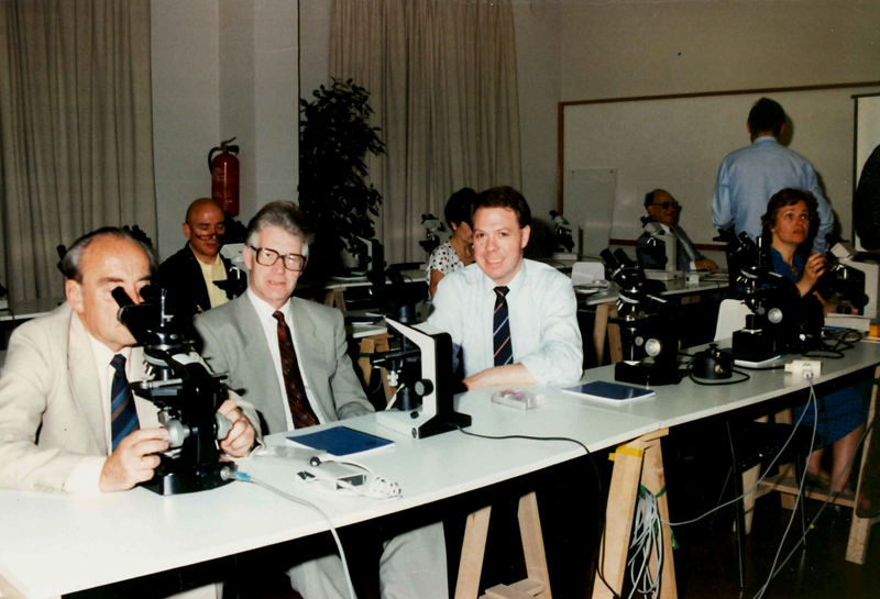
Fig. 3: Professor Dieter Schreiber (second from left) in a seminar in Madrid, 1980. 5.2 Primary tumors of the central nervous system in fetuses and babies Animal experiments showed that tumors in the central nervous system could also be induced transplacentally. This shifted our interest to brain tumors in human fetuses and babies. We derived from studies of the literature documenting that case studies existed, but that systematic examinations on this topic were yet missing. Hence, we initiated a collection and analysis of as much material as possible. The aim was to find out which primary tumors occurred in this age group and which symptoms they exhibited. There were good conditions for this kind of research in the GDR: First, a mandatory national tumor register had been existing since 1952. All physicians were legally obligated to report suspected and evidenced malignant tumors. In 1956, this duty to report was extended to all tumors of the central nervous system, regardless of grade of malignancy. Second, in the GDR there were special consulting centers for all pregnant women and women with babies. Through these, all medical records on the pregnancy and birth including its development and diseases were accessible. Third, autopsy of stillborns and dead babies was mandatory. This way we could access all necessary information and request pathology reports and microscopic preparations. Additionally, our colleagues from abroad reported relevant cases to us and provided us with material to be examined. The support of this endeavor by our colleagues in Poland and Czechoslovakia deserves special thanks. On this basis Dr. Schreiber, Dr. Gerlach and I authored the monography “Tumors of the central nervous system in fetuses and babies” (9). As a supplement to the morphological records, we obtained records on the development of the newborns and babies. We aimed to identify events that might have led to transplacental tumor inductions, and to detect the early symptoms of tumors in the central nervous system of newborns and babies. We intended to include this knowledge into the (continued) training of physicians. We achieved this (13), but the search for transplacental causes for tumors was suspended in 1990. The central tumor register of the GDR was not continued, and with it our access to new data. Further inquiries into the data already collected was foiled by the changes in my professional career, leaving the emerging findings hypothetical and unpublished. 5.3 Epidemiology of primary tumors of the central nervous system The fundamental works on the pathology of tumors of the central nervous system by Zülch (23), Folke Henschen (2), and Russell and Rubinstein (19) give hints on the age and gender distribution of different types of tumors. These are composed of summaries of their own analyses and those of other researchers. However, they do not allow conclusions about the actual frequency in the population; they are not epidemiological findings. An epidemiology of tumors can only be realized if all cases in a predefined population group are recorded over time. Statistics on the material analyzed in some institutions for pathology or some clinics do not amount to epidemiological findings. The existence of the central tumor register of the GDR described above provided us with the opportunity to generate epidemiological findings on primary tumors of the central nervous system in a population of 17 million people. With the help of Dr. Joanna Haas, a well-trained epidemiologist from the U.S., and other staff from the tumor register we were able to generate robust findings. These were published in single publications (5, 10), as well as in books (9, 11). 5.4 Neuroectodermal stem cell tumors Studying tumors of the central nervous system of children sparked a scientific interest in a group of tumors that I summed up using the term “neuroectodermal stem cell tumors.” Hart and Earle (6) called them “primitive neuroectodermal tumors” (PNETs). The term “stem cell tumor” is based on the idea that its cells are at the start of a process that will spawn highly differentiated cells through cell maturation. Such maturation processes can be morphologically demonstrated (15). The cell maturation in stem cell tumors (neuroepithelioma, neuroblastoma, medulloblastoma, medullomyoblastoma etc.) can lead to a decrease of tumor growth and in some cases even to spontaneous recovery of the patients (11, 22). This has been seen with neuroblastoma in the sympathetic nervous system and very rarely also with stem cell tumors in the central nervous system. In many cases, the differentiation process remains on a low level and only gets to part of the tumor tissue, hence having no effect on the course of the disease without improving the prognosis. Furthermore, together with my staff, I intended to research how these maturation processes are controlled, and to find out which therapeutic possibilities might emerge from this. This research produced first results, but had to be suspended in 1994, leaving the intended tasks unfinished. It is my hope that the next generation of researchers will take up this topic and perhaps win the Nobel Prize in medicine.
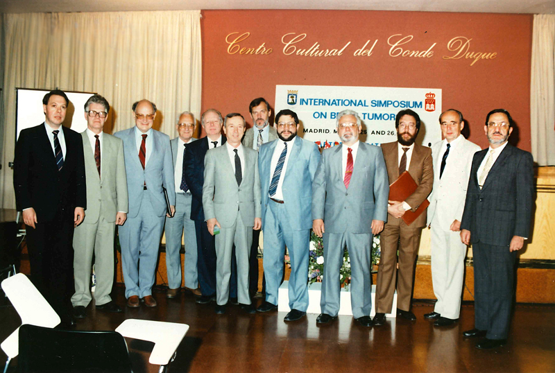
Fig. 4: Participants of the International Symposium on Brain Tumors in Madrid, 1990.
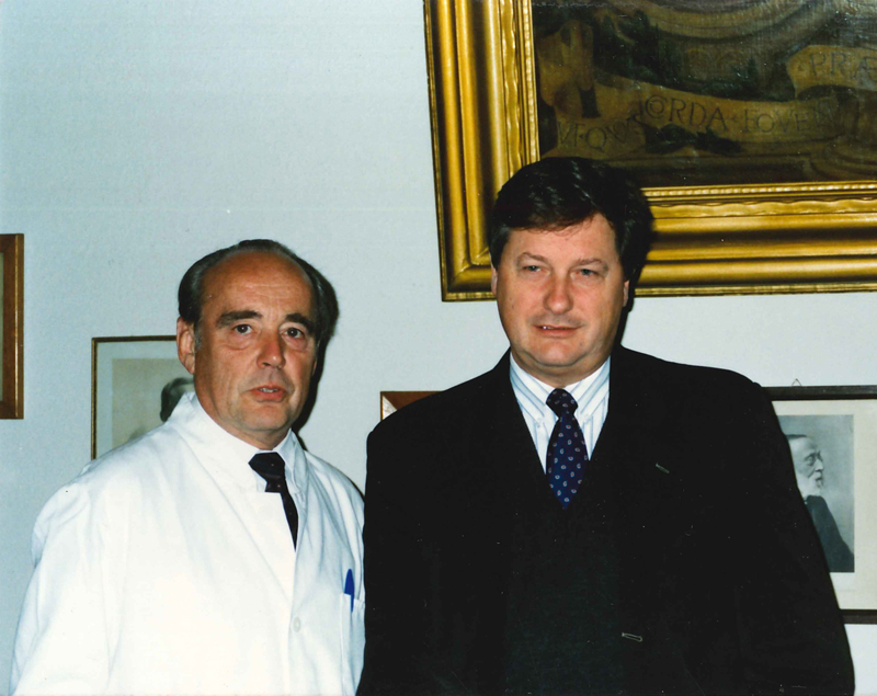
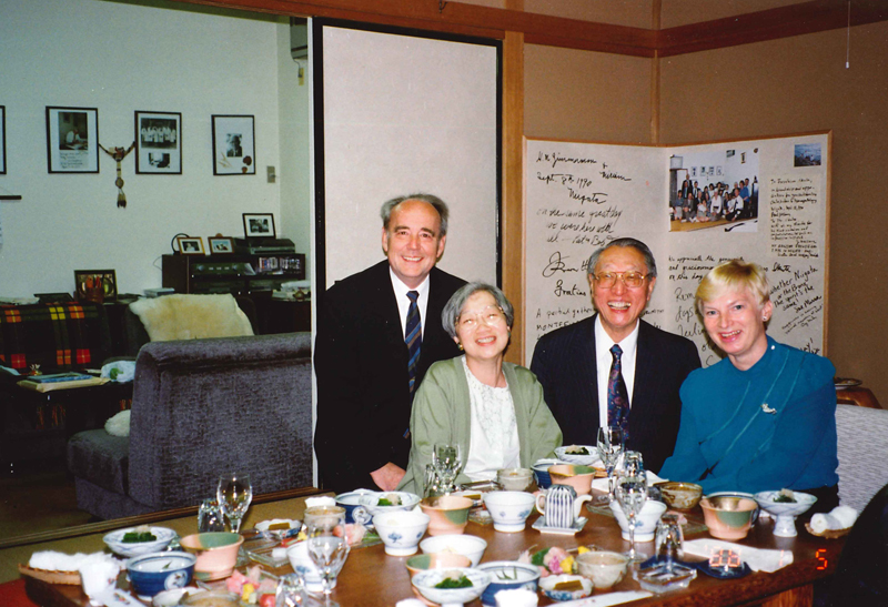
Fig. 5: With Professor Paul Kleihues at the Rudolf Virchow Haus of the Charité in Berlin, 1991. 6. The importance of medical societies and personal contacts It is essential for every physician and professor involved in scientific research to keep up with research findings and the growth of knowledge in the field. This information can be learned from scientific writing, conventions and personal contacts. As a member of the International Society of Neuropathology I took part in several of the meetings and was able to gain many valuable inspirations for my own work; this was also the case for the congresses of the European Confederation of Neuropathological Societies (Euro-CNS). I have been interested in informative presentations and panel discussions, as well as in the opportunity to get to know and discuss specific problems with renowned and next generation researchers from abroad. For example, at the 1970 International Congress of Neuropathology in Paris, I met the well-known veterinary pathologist Professor Fankhauser, co-author of a work on comparative neuropathology (3). I used this opportunity to learn something from him about brain tumors in animals. He then invited me to a multi-week stay in Bern where I had the chance to study his extensive collection of histological preparations of brain tumors of animals. There I also discussed problems of comparative neuropathology with him and Professor Frauchiger. But the exchange of scientific experiences and personal contacts were not limited to international events (Fig. 4); the conventions of national societies of neuropathology and symposia proved valuable, as well. In the GDR, there were the annual conventions of the Society of Neuropathology and symposia with international attendance (20). The proceedings of these meetings were usually published in the journal “Zentralblatt für Allgemeine Pathologie und Pathologische Anatomie” (21). Several of these events were dedicated to tumors of the central nervous system. First they took place in Erfurt, then in Halle/Saale. They offered neuropathologists in the GDR the chance to personally get in touch with researchers from many European countries and countries outside of Europe. Among the frequent guests were Professor Zülch, Professor Wechsler, Professor Kleihues (Fig. 5), Professor Mennel from the Federal Republic of Germany, Professor Cervos-Navarro from West Berlin, Professor Jellinger from Vienna, Professor Mossakowski from Warsaw, and Dr. Jablonowskaja from Moscow, for instance. In exchange, my staff and I were invited to the national neuropathological events in their home countries. Sometimes there were invitations from researchers that I had not met before. Professor Katsuo Ogawa (Fig. 6) from Japan invited me together with Lucy Rorke (Philadelphia) and Umberto Cravioto (New York) to the convention of the Japanese Society of Neuropathology in Okayama in 1985 (Fig. 7). That society had more than 900 members back then (the Society of Neuropathology in the GDR had 34 members.) After that convention I had been invited to talks at different universities and research facilities in Japan, among them Tokyo, Niigata, Maebashi, Nagasaki, and Hiroshima. From this, professional and familial contacts with Japanese pathologists and neuropathologists emerged. In 1992, on the occasion of the congress of neuropathology in Niigata, my wife and I were invited to the house of Professor Ikuta (Fig. 8), where we enjoyed Japanese hospitality together with Professor Hirano (Fig. 9). Then again, my wife and I hosted many peers when they stayed in our hometown (Figures 10 and 11).
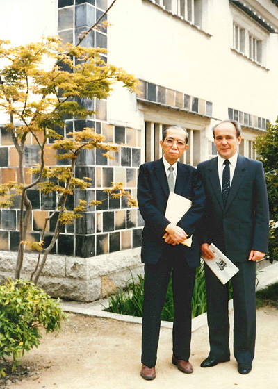
Fig. 6: With Professor Katsuo Ogawa in Okayama, 1985.
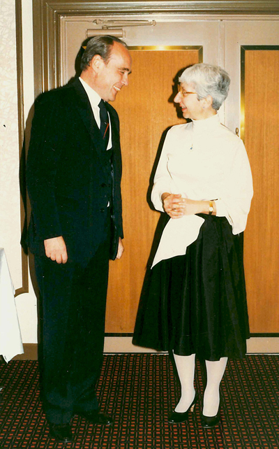
Fig. 7: Discussion with Professor Lucy B. Rorke in Okayama, 1985. 7. Professorship in Iraq In 1967, the government of the Republic of Iraq headed by President Aref asked the government of the GDR to name qualified professors to be temporarily sent to work at Iraqi universities. This was an unusual step, because at this time the GDR had not been internationally acknowledged as a sovereign state. The claim to sole representation by the Federal Republic of Germany (Hallstein doctrine) was still in place. It said that the Federal Republic of Germany is the only successor state of the German Reich and that foreign countries can have diplomatic relations to no other German state. Any violations of this doctrine would be met with extensive sanctions, including the suspension of diplomatic relations and drastic trade restrictions. The only countries excluded from the Hallstein doctrine were those in the socialist realm, which at this point already had the usual diplomatic and other intergovernmental relations with both the GDR and the Federal Republic of Germany in place. The ministry of higher education of the GDR announced the Iraqi government’s request to the country’s professors. I learned about this and expressed my interest in such an activity, however stating that my English skills at that point would not be sufficient for such an appointment. Hence, I was invited to a language course that was offered in small groups for three months under the instruction of experienced simultaneous translators, which turned out to be very effective. In May of 1968, I flew to Baghdad with my wife and kids. We were welcomed by the trade mission of the GDR. I was appointed to act as director of the chair for pathology at the University of Mosul. The trade mission had rented a spacious house on the outskirts of the city, not far from Ninive. Since it was so far away from the medical school, the trade mission provided me with a car of the Soviet brand Lada. The advantage of this car was that it was very sturdy, and therefore always ready to be used in the summer heat and resistant to driving on desert roads. Since this car was the only one of its type in the whole city everyone knew who was coming along when I was going somewhere in it. Despite the lack of any traffic rules, I was never involved in an accident. The department of pathology was housed on the fourth floor of a new building for theoretical medical disciplines and had four large rooms and its own lecture hall for 150 students, but no room for conducting autopsies. The staff consisted of an assistant of Kurdish nationality who had studied veterinary medicine, an English-speaking manager and four male staffers who only spoke in Arabic. One of them was able to prepare paraffin sections and preparations with the usual aniline colorations. The other three assisted by fetching things, cleaning up and making tea. In an adjacent chamber, there was an electron microscope, which had been ordered by one of my predecessors, and it was still unused. There were no tools available for ultrathin sectioning. For the training of the students there were some commercial pathological/anatomical preparations of organs and histological preparations which had been purchased in England. These had been used in classes by the assistant physician. Doing lectures was aided by a blackboard with chalk and a slide projector, but there were no lantern slides. Fortunately, I had brought a large chunk of my collection of diapositives pertaining to general and specific pathology, which could be used to visualize the lectures. Additionally, I demonstrated pathological findings to the students using native organ parts, which came from surgical operations. Among them, echinococcus cysts from the dog tapeworm were frequent, which had been removed by surgeons from various regions of the body such as the liver, kidney and even from the ear lobe. Echinococcus was a frequent disease in Mosul, which was due to the breeding of sheep and the many stray dogs. There were no neurosurgeons active in Mosul. Therefore, I could demonstrate neuropathological findings to the students only in the autopsy room. However, the problem was that patients who had died did not undergo autopsy in the university hospitals. People who had died were usually picked up by their relatives shortly after death and buried before dusk. There was a small, simply equipped autopsy room in an adjacent building on the clinic’s premises, but this had only been used by forensic doctors. These autopsies had been ordered by the court and conducted against the will of the dead person’s relatives. The chair of forensic medicine was held by an Iraqi who had been trained in England. I got in touch with him and we arranged that I would conduct autopsies if the circumstances of the death would allow it. In addition, there was a military hospital in Mosul. They also sometimes requested me to do autopsies. Every time, the building where the autopsy took place was guarded by the military in order to prevent relatives from entering the room, taking the dead person with them, or altercations. It was a problem for me that I could not schedule autopsies for demonstrations with the students, because they had to happen immediately. We solved this problem by sending out my staffers into the clinic building and informing randomly encountered students that there was going to be an autopsy in an hour. This information circulated quickly among the students, leading to crowds in the autopsy room. This way I was able to conduct and analyze more than 20 autopsies together with the students during my work at the University of Mosul. Among them there were heart attacks, fatal lung embolisms, but also neuropathological causes of death, such as hypertensive cerebral hemorrhage or a large tuberculoma in the cerebellum. I would like to stress that the students (nine female and 152 male) were disciplined, curious and grateful.
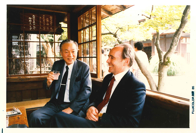
Fig. 8: Break-time talk with Professor Ikuta, director of the Institute for Brain Research at the University of Niigata during the 33rd convention of the Japanese Society of Neuropathology, 1992.
Fig. 9: Staying at Professor Ikuta’s house. Standing between my wife and me: Mrs. Ikuta and Professor Hirano (New York). 8. Deputy Health Minister and WHO consultant Back then, the Minister of Health of the GDR was a physician by the name of Professor Ludwig Mecklinger. He had five deputies. In 1978, the government of the GDR was informed that the Soviet Union would meet its wish to drastically increase oil deliveries. Energy generation through the unprofitable and environmentally harmful combustion of lignite could then be abandoned, leading to massive financial gains for the state. The government, headed by Prime Minister Willy Stoph, planned to allocate a large amount of these gains to medical research. Mecklinger was ordered to lay out plans for this. He asked for a sixth Deputy Health Minister whose main job would be the further development of medical research in the GDR. He wanted somebody who was a scientist himself and was experienced enough to evaluate research endeavors. This is how my name came into play. I declined to take this job because we were making great progress with our research at the department of pathology in Halle. I also stated that I was lacking the necessary expertise in some fields of medicine. I was then assured that a “council for medical research” was to be founded and that renowned scientists from all fields of medicine would constitute this council in order to consult me. This reasoning as well as the significance of medical research for the further development of the health care system of the GDR convinced me to accept the assignment in 1979. I was not influenced by material incentives because even the salary of the Minister of Health of the GDR was below that of a university institute director. We started working, but by the end of 1980 were informed that the Soviet Union would eventually not deliver the amount of oil they had promised before. The power plants, which in the meantime had been adapted to the combustion of oil, had to be quickly reconfigured back to the combustion of lignite. This cost a lot of money, leading to not more but less investments in medical research. Mecklinger was a smart and warmhearted person. He accepted that my position was not necessary anymore and let me leave the Ministry in order to return to the university. This was how I came to the Charité Berlin in 1983. The “council for medical research” continued to exist and I belonged to it until its dissolution by the last Minister of Health, Professor Kleditzsch, in 1990. From 1986 to 1990, I was also acting as consultant to the WHO, where I was involved in the revision of the International Classification of Diseases (ICD-10). Therefore, I spent multiple months per year in Geneva. This way I gained insight into the way a United Nations agency works and got to know many professionals from several countries, which allowed me to expand my worldview. 9. The end of my academic career and research activities The integration of the GDR into the Federal Republic of Germany brought along changes for many citizens of the GDR, and for me as well. In March of 1991 I was told that the senate administration for internal affairs in West Berlin had ordered a vetting of all employees working in the public sector in East Berlin (formerly GDR). It was to be decided who would be able to transition into the public sector of the Federal Republic of Germany and who would have to be let go. The focus was on personal aptitude, and on whether or not somebody had worked for the Ministry of State Security (MfS). This also concerned the professors of the Humboldt University. Hence, I was asked to answer an extensive questionnaire. On August 27, 1991 I was informed that “no indications of formal or informal activities for the MfS were found and that for this reason no measures concerning employment law [were] to be taken.” I was allowed to continue my activities in the training of students, supervision of graduates and doctoral students, diagnostics of biopsies and autopsies, and heading the department of neuropathology. Based on provisions in the Unification Treaty I was allowed to continue to bear the title “full professor” (“ordentlicher Professor”), which I was awarded in 1970 by the ministry of higher education of the GDR. However, the title did not imply an according salary. In the Federal Republic of Germany, professors were paid according to the salary groups “C3” or “C4”. My salary group equaled that of an assistant physician. It was my ethical belief as a professional that I would continue teaching and do my work in neuropathology until my retirement regardless of my salary. However, I was later informed that I would be able to apply as a professor. My position was advertised for applicants from the whole country under the heading “C3 professorship of pathology with emphasis on neuropathology.” I was allowed to apply to that, as well. I and four other applicants from West Berlin and West Germany were asked to give a talk in front of a commission. After that, we were interviewed in person. During my interview, the chairman of the commission, Professor Detlev Ganden, was especially interested in my research on neuroectodermal stem cell tumors. I have never been notified about the result of the selection process. On December 2, 1993 I was notified by the personnel office of the Charité that the selection process became unnecessary “because the advertised position was not part of the legally binding employment plan of Humboldt University.” In the same letter, I was told that I would be terminated effective February 2, 1994, because “there [were] concerns regarding my personal aptitude for continued employment in the public sector.” I only received this letter after returning from vacation in Indonesia that I had spent with my wife from December 1–22, 1993. I refrained from filing a lawsuit because I believed (and still do believe) that I would not force myself on anybody if my special knowledge and my efforts in the (continued) training of the next generation of physicians was not deemed necessary. I have explained this opinion to the institute’s staff when they tried to encourage me to take steps pertaining to employment law. Apparently, the administrative director of the Charité heard about this, too, because in mid-January of 1994 he offered me an annulment contract instead of a termination if I refrained from suing them. The benefit was that the contract would not be effective February 2, but February 28. This was indeed of great significance to me, because I had been storing large collections of scientific literature, photographic documentations, case studies and correspondence (I was still editor of the Zentralblatt) at the institute. Because I was given more time, I was able to rent a large storage facility, which my son helped me to store all the irreplaceable material in. We saved it from destruction, because the newly appointed director of the institute threatened to order the removing of everything in the building as soon as I had been dismissed. I was surprised that the annulment contract did not mention the “insufficient personal aptitude” as a reason for the threatened termination.
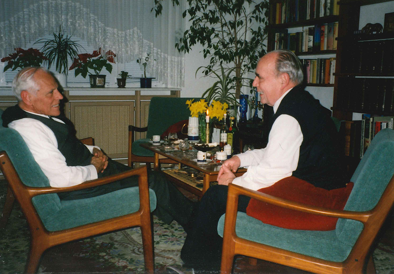
Fig. 10: Professor Jorge Cervos-Navarro, director of the Institute of Neuropathology at Free University Berlin, at our apartment, 2002.
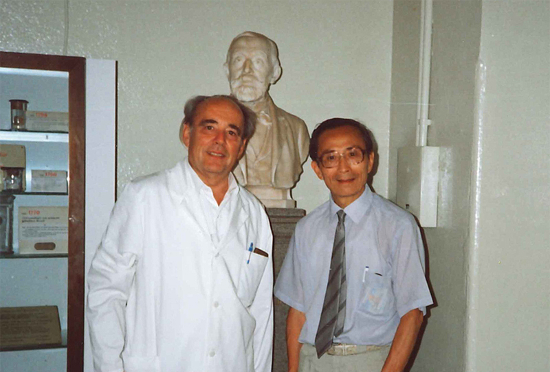
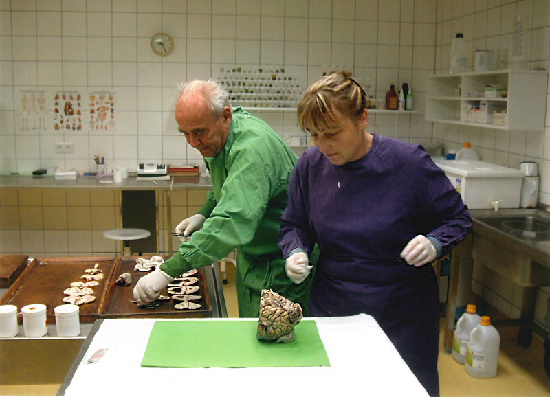
Fig. 11: Visitor from East Asia in the Rudolf Virchow House of the Charité Berlin, 1992 (Note the Virchow bust in the background). 10. Restarting my career Shortly after the announcement of my separation from the Charité the medical director of the Landesklinik Brandenburg offered me to become director of their department of neuropathology. It had five staffers, was well-equipped and had its own electron microscope (Fig. 12). My main tasks would be to examine the brains of patients who had died (autopsies would be conducted at the Institute of Pathology of the Municipal Clinic Brandenburg) and examining nerve and muscle biopsies. In addition, I was told that in a few months neurosurgery would be opened at the Municipal Clinic Brandenburg and that we would be given tissue samples for examination. This was the reason for me to accept the job offer. Then the Prime Minister of Brandenburg converted the position of the head of department into that of a chief physician, thus putting it on the same level with the chief physicians of the clinics for neurology and psychiatry. One of the staffers in the department was Dr. Hermann, who had special skills in the area of morphological diagnostics of muscle and nerve biopsies. He undertook this part of the department’s tasks, which was good for me, as I did not have deep knowledge of this special area. Working in this department and the cooperation between the clinics was good. Clinicopathological conferences were introduced and occurred on a regular basis. Physicians from the Landesklinik as well as the Municipal Clinic for Neurosurgery participated. The exchange of opinion that took place furthered the cooperation of the different disciplines to the advantage of the patients. Since the chief physicians participated themselves in these conferences, they served as continued training for all participants. There also emerged a close cooperation with the Brandenburg State Institute for Forensic Medicine in Potsdam. The forensic doctors asked for our help when examinations of the brain and spinal cord of patients who had died were of forensic significance. We were given these organs in toto, sectioned them, and photographically documented macroscopic findings. After microscopic examinations were finished, the forensic doctors received a detailed report of the findings, which included an epicritic evaluation. We were never present in court ourselves and only rarely corresponded with the prosecution. We also had neuropathological conferences involving the forensic doctors. When I reached retirement age the director of the Landesklinik dismissed me, but shortly after Dr. Pauli, chief physician of the Pathological Institute at the Municipal Clinic Brandenburg, offered me to work with him as a pathologist and neuropathologist. This way the cooperation with the neurosurgeons and forensic doctors could be continued. Back when I was working at the Landesklinik, an assistant at the Institute for Pathology in Brandenburg who was in special training, Dr. Marlies Günther (Fig. 13), asked me to help her become acquainted with the field of neuropathology. Hence, we could continue our cooperation until the point where she could conduct neuropathological diagnostics herself. I took pleasure in this cooperation because of her thirst for knowledge. My presence at the institute in Brandenburg was usually limited to three days per week. Therefore, it was possible for me to accept an offer to work at the Institute of Neuropathology at Free University Medical School Berlin. The dean asked for my temporary help because the former director of the institute, Professor Cervos-Navarro, had left after reaching retirement age, and his replacement, Professor Gisela Stoltenburg-Didinger, was on sick leave. I agreed to help on two days per week. The job was mainly concerned with the diagnostics of neurosurgical material and postmortal neuropathology. I was not asked to be involved with the training of students. A good cooperation with the director of neurosurgery, Prof. Brock, and his staffers quickly emerged. Intraoperative frozen sections which were prepared in a room next to the operating room and diagnosed by me, fostered this cooperation. This way I was able to communicate and discuss the diagnosis with the surgeon instantly. He then demonstrated the intraoperative situs to me on a display and told me details that would be relevant for my decisions.
Fig. 12: Working in the Department of Neuropathology of the Landesklinik Brandenburg, 1995.
Fig. 13: Working with Dr. Marlies Günther. 11. As a neuropathologist in Thailand After my dismissal by the directors of the Charité, I continued to receive invitations for conventions in the field of neuropathology. One of them became a special memory: It came from the medical school of the University in Khon Kaen in Northeastern Thailand. I was asked to organize a workshop for physicians consisting of presentations, autopsy seminars and case study discussions about tumors, inflammations and parasitoses in the central nervous system over the span of several days. The discussions were supposed to give participants the opportunity to present cases of their own choosing, request my opinion on them and discuss them together. I was also asked to give a lecture on neuropathology and answer medical students’ questions. The expenses of this ten-day journey were covered by a Japanese sponsor of the university. The offer was appealing, but also somewhat risky as I did not have any information on how knowledgeable the participants would be, what their cases might be about, and if the students would even be interested in neuropathology. Furthermore, the events was planned to take place in English and I was not sure if my presentations would be comprehensible. Therefore, I prepared for the events using my extensive collection of diapositives because my previous teaching activity in Mosul showed to me that language barriers could be overcome using illustrative and expressive diapositives. About 60 physicians took part with the workshops I offered for postgraduates. The physicians and students were attentive and engaged in sober discussions. The lecture hall was filled with students up to the last seat. My wife took a seat in the last row and later reported to me how attentively everyone was listening to my delineations. I felt this myself when, after a one-hour lecture, I invited listeners to ask questions. For more than two hours, I replied to profound inquiries, which were proof of the good knowledge basis and intellect of the Thai students. This experience was of great joy to me and made the extensive preparations for these events pay off. 12. Epilogue My activities in neuropathology teaching and research came to an abrupt halt in the spring of 1994. In 2012, I decided to conclude my activities as a pathologist and neuropathologist. As of this writing, I am living in the 89th year of my life. If someone asked me if would choose this career again, I would clearly reply “Yes!” However, I have to advise younger readers that they cannot expect material wealth from working in this area. I am not the only person who had to make the experience of working for an hourly wage of two or three cents (co-)authoring neuropathological books (11, 12). I emphasized this in the prologue: This is not an autobiography. However, I still would like to briefly express my commitment to my philosophy of life: My childhood experiences made me a staunch enemy of National Socialist ways of governing and the horrors of World War II made me an opponent of war. I did not enroll in the army. The experiences of the Japanese people taught me that nuclear armament poses a threat to civilization. Therefore I joined the organization “International Physicians for the Prevention of Nuclear War (IPPNW)” in 1980 and worked for their cause. This worldview determines the way I live until this day. References 1. Bailey P., Cushing H.: Gewebsverschiedenheit der Hirngliome. Gustav Fischer-Verlag, Jena 1930 2. Henschen, F.: Tumoren des Zentralnervensystems und seiner Hüllen. Handbuch der speziellen pathologischen Anatomie und Histologie, herausgeg von O. Lubarsch, F. Henke, R.Rössle. Band 13, Teil 3; Springer-Verlag, Berlin-Göttingen-Heidelberg, 1955 3. Frauchiger E., Fankhhauser R.: Vergleichende Neuropathologie des Menschen und der Tiere. Springer-Verlag, Berlin-Göttingen-Heidelberg 1957 4. Gerlach H., Jänisch W., Schreiber D.: Perinatale Teratome des Zentralnervensystems. Wiss. Z. Ernst-Moritz-Arndt-Univ. Greifswald 32: 112-114; 1983 5. Haas J.F., Jänisch W., Staneczek W.: Newly diagnosed primary intracranial neoplasms in pregnant woman: a population-based assessment. J. Neurol. Neurosurg. Psychiat. 49: 874-800; 1986 6. Hart M.N., Earle K.M.: Primitive neuroectodermal tumors of the brain in children. Cancer 32: 890.897; 1973 7. Jänisch W., Schreiber D.: Experimentelle Geschwülste des Zentralnervensystems. VEB Gustav Fischer-Verlag, Jena 1969 8. Jänisch W., Schreiber D.: Experimental tumors of the central nervous system. Edited by D.D.Bigner and J. Swenberg; Upjohn Comp., Kalamazoo/Mich.; 1977 9. Jänisch W., Schreiber D., Gerlach H.: Tumoren des Zentralnervensystems bei Feten und Säuglingen. VEB Gustav Fischer-Verlag, Jena 1980 10. Jänisch W.: Zur Epidemiologie der primären Geschwülste des Zentralnervensystems im ersten Lebensjahr. Arch. Geschwulstforsch. 55: 489-494; 1985 11. Jänisch W., Schreiber D., Güthert H.: Neuropathologie (Band 1), Tumoren des Nervensystems. VEB Gustav Fischer-Verlag, Jena 1988 12. Jänisch W., Schreiber D., Warzok R.: Neuropathologie (Band 2), Pathomorphologie und Pathogenese neurologischer Krankheiten. VEB Gustav Fischer-Verlag, Jena 1990 13. Jänisch W.: Hirngeschwülste bei Säuglingen. Z. Allg. Med. 65: 484-489; 1989 14. Jänisch W: Pathologie der Geschwülste des Nervensystems. In: Klinische Neuropathologie. Herausgegeben von J. Cervos-Navarro u. R. Ferszt: Georg Thieme-Verlag, Stuttgart-New York; 1989 15. Jänisch W., Grieshammer T.: Expression von immunhistochemischen Differenzierungsmarkern i n normalen und neoplastisch transformierten neuroektodermalen Stammzellen. Acta histochemica Suppl.-Band XLII: 139-142; 1992 16. Jänisch W.: Nocardiose. In: Die entzündlichen Erkrankungen des Nervensystems, Band 2; herausgeg. von H. Henkes u. H.W. Kölmel; Ecomed-Verlag, Landsberg/Lech 1993 17. Jänisch W.: Mykosen des Zentralnervensystems. In: Die entzündlichen Erkrankungen des Nervensystems, Band 5; herausgeg. von H. Henkes u. H.W. Kölmel; Ecomed-Verlag, Landsberg/Lech 1993 18. Paulus W., Jänisch W.: Clinicopathologic correlations in epithelial choroid plexus neoplasms: a study of 52 cases. Acta Neuropathol. 80: 635-641; 1990 19. Russell D.S., Rubinstein L.J.: Pathology of Tumours of the Nervous System. Edward Arnold , London 1959 20. Schreiber D. und Jänisch W. (Herausgeber): Experimentelle Neuroonkologie. Wissenschaftliche Beiträge der Martin-Luther –Universität Halle-Wittenberg; Heft 7, 1974 21. Schreiber D., Güthert H.: Bericht über die 2. Tagung der Gesellschaft für Neuropathologie der DDR am 1. Und 2. Oktober 1970 in Erfurt. Zbl. allg. Path. 114: 276-285, 1971 22. Warzok R., Jänisch W., Lang G.: Morphology and biology of cerebellar neuroblastomas. J. Neuro-Oncol. 1: 373-379; 1983 23. Zülch K.J.: Die Hirngeschwülste in biologischer und morphologischer Darstellung. Barth-Verlag, Leipzig 1956
Copyright: © 2020 The author(s). This is an open access article distributed under the terms of the Creative Commons Attribution 4.0 International License (https://creativecommons.org/licenses/by/4.0/), which permits unrestricted use, distribution, and reproduction in any medium, provided the original author and source are credited, a link to the Creative Commons license is provided, and any changes are indicated. The Creative Commons Public Domain Dedication waiver (https://creativecommons.org/publicdomain/zero/1.0/) applies to the data made available in this article, unless otherwise stated. |